Department of Cardio-Thoracic Surgery, University Medical Center, Utrecht, The Netherlands.
J Cell Mol Med. 2012 Apr;16(4):730-9. doi: 10.1111/j.1582-4934.2011.01351.x.
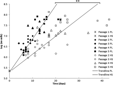
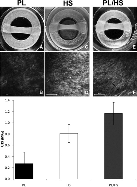
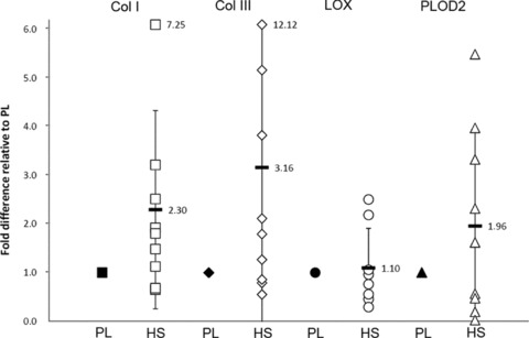
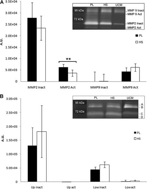
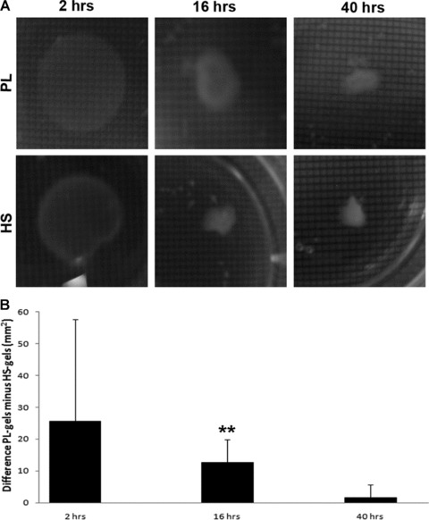
For clinical application of tissue engineering strategies, the use of animal-derived serum in culture medium is not recommended, because it can evoke immune responses in patients. We previously observed that human platelet-lysate (PL) is favourable for cell expansion, but generates weaker tissue as compared to culture in foetal bovine serum (FBS). We investigated if human serum (HS) is a better human supplement to increase tissue strength. Cells were isolated from venous grafts of 10 patients and expanded in media supplemented with PL or HS, to determine proliferation rates and expression of genes related to collagen production and maturation. Zymography was used to assess protease expression. Collagen contraction assays were used as a two-dimensional (2D) model for matrix contraction. As a prove of principle, 3D tissue culture and tensile testing was performed for two patients, to determine tissue strength. Cell proliferation was lower in HS-supplemented medium than in PL medium. The HS cells produced less active matrix metallo-proteinase 2 (MMP2) and showed increased matrix contraction as indicated by gel contraction assays and 3D-tissue culture. Tensile testing showed increased strength for tissues cultured in HS when compared to PL. This effect was more pronounced if cells were sequentially cultured in PL, followed by tissue culture in HS. These data suggest that sequential use of PL and HS as substitutes for FBS in culture medium for cardiovascular tissue engineering results in improved cell proliferation and tissue mechanical properties, as compared to use of PL or HS apart.
对于组织工程策略的临床应用,不建议在培养基中使用动物源性血清,因为它会在患者中引发免疫反应。我们之前观察到,人血小板裂解物(PL)有利于细胞扩增,但与胎牛血清(FBS)培养相比,生成的组织较弱。我们研究了人血清(HS)是否是一种更好的人类补充物,可以增加组织强度。从 10 名患者的静脉移植物中分离细胞,并在添加 PL 或 HS 的培养基中进行扩增,以确定增殖率和与胶原产生和成熟相关的基因表达。通过明胶酶谱法评估蛋白酶表达。胶原收缩测定法用于评估基质收缩的二维(2D)模型。作为原理验证,对两名患者进行了 3D 组织培养和拉伸测试,以确定组织强度。HS 补充培养基中的细胞增殖低于 PL 培养基。HS 细胞产生的活性基质金属蛋白酶 2(MMP2)较少,通过凝胶收缩测定和 3D 组织培养表明基质收缩增加。与 PL 相比,HS 培养的组织拉伸测试显示出更高的强度。如果细胞依次在 PL 中培养,然后在 HS 中进行组织培养,则效果更为明显。这些数据表明,与单独使用 PL 或 HS 相比,在培养基中顺序使用 PL 和 HS 作为心血管组织工程中 FBS 的替代品可提高细胞增殖和组织力学性能。