Cardiovascular Research Center, Mount Sinai School of Medicine, New York, New York, United States of America.
PLoS One. 2011 Apr 20;6(4):e19097. doi: 10.1371/journal.pone.0019097.
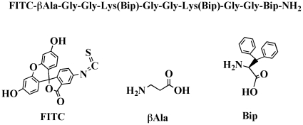
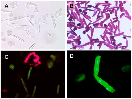
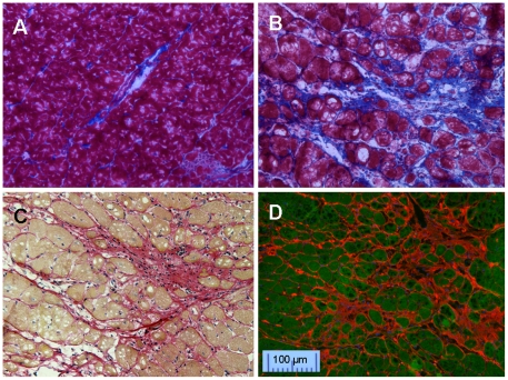
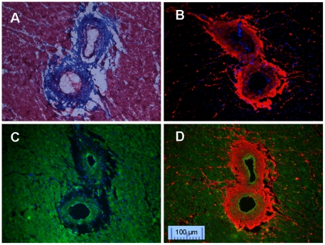
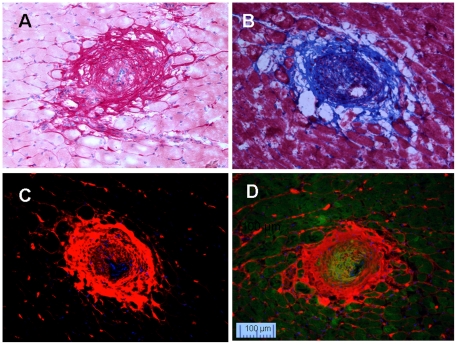
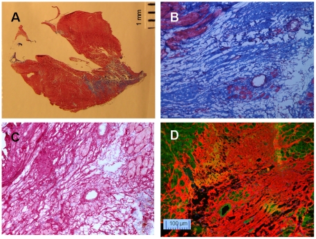
Cardiovascular fibrosis resulted from pressure overload or ischemia could alter myocardial stiffness and lead to ventricular dysfunction. Fluorescently labeled collagen-binding protein CNA 35, derived from the surface component of Staphylococcus aureus, and a novel synthetic biphenylalanine containing peptide are applied to stain fibrosis associated collagen and myocytes, respectively. Detailed pathological characteristics of cardiovascular fibrosis could be identified clearly in 2 hours. This staining pair requires only simple staining and brief washing, generating less than 10 ml of waste. The image information collected by this novel fluorescent staining pair is compatible with it collected by the traditional Masson's Trichrome and Picrosirius Red staining which are widely used to stain cardiovascular fibrosis and isolated cells.
心血管纤维化是由压力超负荷或缺血引起的,可改变心肌硬度,导致心室功能障碍。荧光标记的胶原结合蛋白 CNA35 来源于金黄色葡萄球菌表面成分,一种新型合成的含二苯丙氨酸的肽分别用于染色纤维化相关的胶原和心肌细胞。在 2 小时内可以清楚地识别心血管纤维化的详细病理特征。这种染色对仅需要简单染色和短暂洗涤,产生的废物少于 10 毫升。这种新型荧光染色对收集的图像信息与广泛用于染色心血管纤维化和分离细胞的传统 Masson 三色和苦味酸红染色收集的信息兼容。