• 文献检索
  • 文档翻译
  • 深度研究
  • 学术资讯
  • Suppr Zotero 插件Zotero 插件
  • 邀请有礼
  • 套餐&价格
  • 历史记录
应用&插件
Suppr Zotero 插件Zotero 插件浏览器插件Mac 客户端Windows 客户端微信小程序
定价
高级版会员购买积分包购买API积分包
服务
文献检索文档翻译深度研究API 文档MCP 服务
关于我们
关于 Suppr公司介绍联系我们用户协议隐私条款
关注我们

Suppr 超能文献

核心技术专利:CN118964589B侵权必究
粤ICP备2023148730 号-1Suppr @ 2026

文献检索

告别复杂PubMed语法,用中文像聊天一样搜索,搜遍4000万医学文献。AI智能推荐,让科研检索更轻松。

立即免费搜索

文件翻译

保留排版,准确专业,支持PDF/Word/PPT等文件格式,支持 12+语言互译。

免费翻译文档

深度研究

AI帮你快速写综述,25分钟生成高质量综述,智能提取关键信息,辅助科研写作。

立即免费体验

1.5T 和 3T MRI 扫描仪在评估足部急性骨应力中的比较。

Comparison of 1.5T and 3T MRI scanners in evaluation of acute bone stress in the foot.

机构信息

Research Department, Centre for Military Medicine, P,O, Box 50, FIN-00301 Helsinki, Finland.

出版信息

BMC Musculoskelet Disord. 2011 Jun 6;12:128. doi: 10.1186/1471-2474-12-128.

DOI:10.1186/1471-2474-12-128
PMID:21645348
原文链接:https://pmc.ncbi.nlm.nih.gov/articles/PMC3121660/
Abstract

BACKGROUND

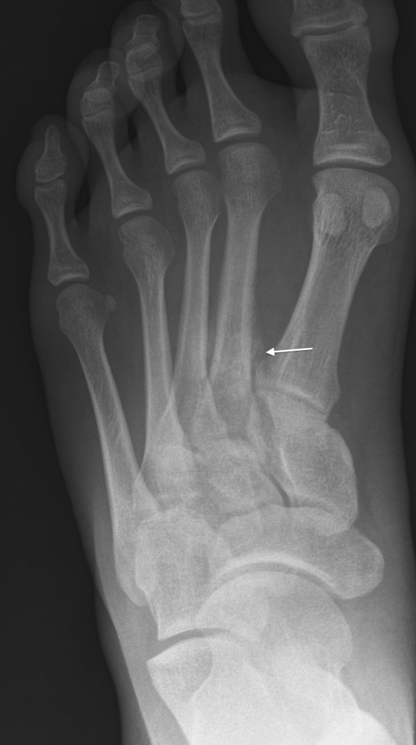
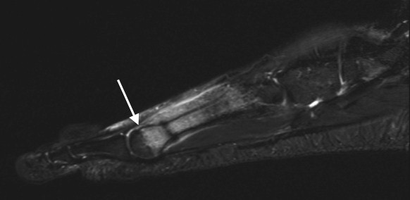
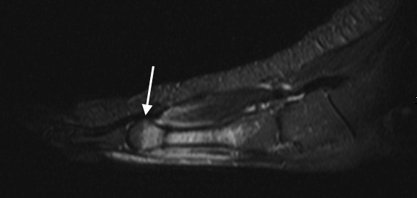
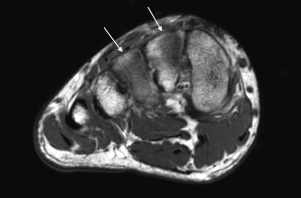
Bone stress injuries are common in athletes and military recruits. Only a minority of bone stress changes are available on plain radiographs. Acute bone stress is often visible on MRI as bone marrow edema, which is also seen in many other disease processes such as malignancies, inflammatory conditions and infections. The purpose of this study was to investigate the ability of radiographs, 1.5T and 3T MRI to identify acute bone marrow changes in the foot.

METHODS

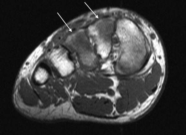
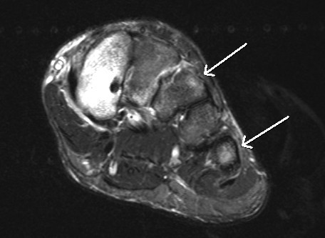
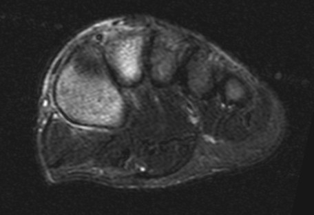
Ten patients with 12 stress fractures seen on plain radiographs underwent MRI using 1.5T and 3T scanners. T1 FSE and STIR axial, sagittal, and coronal view sequences were obtained. Two musculoskeletal radiologists interpreted the images independently and by consensus in case of disagreement.

RESULTS

Of the 63 acute bone stress changes seen on 3T images, 61 were also seen on 1.5T images. The sensitivity of 1.5T MRI was 97% (95% CI: 89%-99%) compared with 3T. The 3T MRI images where, therefore, at least equally sensitive to 1.5T scanners in detection of bone marrow edema. On T1-weighted sequences, 3T images were slightly superior to 1.5T images in visualizing the demarcation of the edema and bone trabeculae. The kappa-value for inter-observer variability was 0.86 in the MRI indicating substantial interobserver agreement.

CONCLUSIONS

Owing to slightly better resolution of 3T images, edema characterization is easier, which might aid in the differential diagnosis of the bone marrow edema. There was, however, no noteworthy difference in the sensitivity of the 1.5T and 3T images to bone marrow edema. Routine identification of acute bone stress changes and suspected stress injuries can, therefore, be made with 1.5T field strength.

摘要

背景

骨应力损伤在运动员和新兵中很常见。只有少数骨应力变化可在普通 X 光片上看到。急性骨应力在 MRI 上通常表现为骨髓水肿,这种情况也可见于许多其他疾病过程,如恶性肿瘤、炎症性疾病和感染。本研究旨在探讨 X 线、1.5T 和 3T MRI 识别足部急性骨髓变化的能力。

方法

10 例在普通 X 光片上发现的 12 例应力性骨折患者接受了 1.5T 和 3T 扫描仪的 MRI 检查。获得了 T1 FSE 和 STIR 轴位、矢状位和冠状位图像。两位肌肉骨骼放射科医生独立解读图像,如果意见不一致,则通过共识进行解读。

结果

在 3T 图像上看到的 63 个急性骨应力变化中,有 61 个也在 1.5T 图像上看到。1.5T MRI 的敏感性为 97%(95%CI:89%-99%),与 3T 相比。因此,在检测骨髓水肿方面,3T MRI 图像与 1.5T 扫描仪的敏感性至少相当。在 T1 加权序列上,3T 图像在显示水肿和骨小梁的边界方面略优于 1.5T 图像。MRI 中观察者间变异的 Kappa 值为 0.86,表明观察者间存在实质性一致性。

结论

由于 3T 图像的分辨率略高,因此更容易对水肿进行特征描述,这可能有助于骨髓水肿的鉴别诊断。然而,1.5T 和 3T 图像对骨髓水肿的敏感性没有明显差异。因此,可以使用 1.5T 场强来常规识别急性骨应力变化和疑似应力性损伤。

https://cdn.ncbi.nlm.nih.gov/pmc/blobs/a258/3121660/e41638d67655/1471-2474-12-128-7.jpg
https://cdn.ncbi.nlm.nih.gov/pmc/blobs/a258/3121660/067dcd5a4faa/1471-2474-12-128-1.jpg
https://cdn.ncbi.nlm.nih.gov/pmc/blobs/a258/3121660/265150c6f6bf/1471-2474-12-128-2.jpg
https://cdn.ncbi.nlm.nih.gov/pmc/blobs/a258/3121660/7be17485d1f6/1471-2474-12-128-3.jpg
https://cdn.ncbi.nlm.nih.gov/pmc/blobs/a258/3121660/ed5a0f8a6a19/1471-2474-12-128-4.jpg
https://cdn.ncbi.nlm.nih.gov/pmc/blobs/a258/3121660/2e030c30c43c/1471-2474-12-128-5.jpg
https://cdn.ncbi.nlm.nih.gov/pmc/blobs/a258/3121660/142a167a6f69/1471-2474-12-128-6.jpg
https://cdn.ncbi.nlm.nih.gov/pmc/blobs/a258/3121660/e41638d67655/1471-2474-12-128-7.jpg

相似文献

1
Comparison of 1.5T and 3T MRI scanners in evaluation of acute bone stress in the foot.1.5T 和 3T MRI 扫描仪在评估足部急性骨应力中的比较。
BMC Musculoskelet Disord. 2011 Jun 6;12:128. doi: 10.1186/1471-2474-12-128.
2
MR imaging of fatigue stress injuries to bones: intra- and interobserver agreement.骨骼疲劳性应力损伤的磁共振成像:观察者间及观察者内一致性
Magn Reson Imaging. 2002 Jun;20(5):401-6. doi: 10.1016/s0730-725x(02)00514-3.
3
Bone stress injuries of the ankle and foot: an 86-month magnetic resonance imaging-based study of physically active young adults.踝关节和足部的骨应力损伤:一项基于磁共振成像的对身体活跃的年轻成年人进行的86个月研究。
Am J Sports Med. 2007 Apr;35(4):643-9. doi: 10.1177/0363546506295701. Epub 2006 Nov 12.
4
Bone marrow abnormalities of foot and ankle: STIR versus T1-weighted contrast-enhanced fat-suppressed spin-echo MR imaging.足踝部骨髓异常:短T1反转恢复序列(STIR)与T1加权对比增强脂肪抑制自旋回波磁共振成像对比
Radiology. 2002 Aug;224(2):463-9. doi: 10.1148/radiol.2242011252.
5
Bone marrow oedema assessment by magnetic resonance imaging in rheumatoid arthritis wrist and metacarpophalangeal joints: the importance of field strength, coil type and image resolution.磁共振成像评估类风湿关节炎腕关节和掌指关节骨髓水肿:场强、线圈类型和图像分辨率的重要性。
Rheumatology (Oxford). 2014 Aug;53(8):1446-51. doi: 10.1093/rheumatology/keu029. Epub 2014 Mar 21.
6
Stress injuries of the calcaneus detected with magnetic resonance imaging in military recruits.新兵中通过磁共振成像检测到的跟骨应力性损伤。
J Bone Joint Surg Am. 2006 Oct;88(10):2237-42. doi: 10.2106/JBJS.E.01447.
7
Comparative reliability and diagnostic performance of conventional 3T magnetic resonance imaging and 1.5T magnetic resonance arthrography for the evaluation of internal derangement of the hip.比较常规 3T 磁共振成像和 1.5T 磁共振关节造影在评估髋关节内部紊乱方面的可靠性和诊断性能。
Eur Radiol. 2018 Mar;28(3):963-971. doi: 10.1007/s00330-017-5069-4. Epub 2017 Oct 6.
8
Deep learning reconstruction for the evaluation of neuroforaminal stenosis using 1.5T cervical spine MRI: comparison with 3T MRI without deep learning reconstruction.使用 1.5T 颈椎 MRI 进行神经孔狭窄的深度学习重建评估:与无深度学习重建的 3T MRI 的比较。
Neuroradiology. 2022 Oct;64(10):2077-2083. doi: 10.1007/s00234-022-03024-6. Epub 2022 Aug 3.
9
[Magnetic-resonance imaging in the diagnosis of overload injuries of bones].[磁共振成像在骨骼过载损伤诊断中的应用]
Lik Sprava. 2003 Jul-Aug(5-6):64-7.
10
Single-Shot Echo-Planar Diffusion-Weighted MR Imaging at 3T and 1.5T for Differentiation of Benign Vertebral Fracture Edema and Tumor Infiltration.3T和1.5T单次激发回波平面扩散加权磁共振成像用于鉴别良性椎体骨折水肿和肿瘤浸润
Korean J Radiol. 2016 Sep-Oct;17(5):590-7. doi: 10.3348/kjr.2016.17.5.590. Epub 2016 Aug 23.

引用本文的文献

1
A method for sensitivity analysis of automatic contouring algorithms across different contrast weightings using synthetic magnetic resonance imaging.一种使用合成磁共振成像对不同对比度加权下的自动轮廓算法进行敏感性分析的方法。
Phys Imaging Radiat Oncol. 2025 May 28;35:100790. doi: 10.1016/j.phro.2025.100790. eCollection 2025 Jul.
2
A Method for Sensitivity Analysis of Automatic Contouring Algorithms Across Different MRI Contrast Weightings Using SyntheticMR.一种使用SyntheticMR对不同MRI对比加权下自动轮廓算法进行敏感性分析的方法。
medRxiv. 2025 Jan 12:2025.01.10.25319895. doi: 10.1101/2025.01.10.25319895.
3

本文引用的文献

1
Bone stress injury of the ankle in professional ballet dancers seen on MRI.磁共振成像显示的职业芭蕾舞演员踝关节骨应力损伤
BMC Musculoskelet Disord. 2008 Mar 28;9:39. doi: 10.1186/1471-2474-9-39.
2
Fast high-spatial-resolution MRI of the ankle with parallel imaging using GRAPPA at 3 T.3T下使用GRAPPA并行成像的踝关节快速高空间分辨率MRI
AJR Am J Roentgenol. 2007 Jul;189(1):240-5. doi: 10.2214/AJR.07.2066.
3
Three-tesla MR imaging of the knee.膝关节的3特斯拉磁共振成像。
Bone age assessment based on different MRI modalities of the proximal humerus epiphysis: the comparisons of TWI, TWI, and PDWI.
基于肱骨近端骨骺不同 MRI 模式的骨龄评估:TWI、TWI 和 PDWI 的比较。
Int J Legal Med. 2024 Jul;138(4):1509-1521. doi: 10.1007/s00414-024-03182-7. Epub 2024 Feb 9.
4
Towards a simplified fluid-sensitive MRI protocol in small joints of the hand in early arthritis patients: reliability between modified Dixon and regular Gadolinium enhanced TSE fat saturated MRI-sequences.针对早期关节炎患者手部小关节简化液体敏感 MRI 方案:改良 Dixon 与常规钆增强 TSE 脂肪饱和 MRI 序列之间的可靠性。
Skeletal Radiol. 2023 Jun;52(6):1193-1202. doi: 10.1007/s00256-022-04238-8. Epub 2022 Nov 28.
5
Rib Fractures in Professional Baseball Pitchers: Mechanics, Epidemiology, and Management.职业棒球投手的肋骨骨折:力学、流行病学与管理
Open Access J Sports Med. 2022 Oct 10;13:89-105. doi: 10.2147/OAJSM.S288882. eCollection 2022.
6
The Effect of Magnetic Field Gradient and Gadolinium-Based MRI Contrast Agent Dotarem on Mouse Macrophages.磁场梯度和基于钆的 MRI 造影剂 Dotarem 对小鼠巨噬细胞的影响。
Cells. 2022 Feb 22;11(5):757. doi: 10.3390/cells11050757.
7
TREAT Early Arthralgia to Reverse or Limit Impending Exacerbation to Rheumatoid arthritis (TREAT EARLIER): a randomized, double-blind, placebo-controlled clinical trial protocol.早期关节炎治疗以逆转或限制类风湿关节炎恶化(更早治疗关节炎):一项随机、双盲、安慰剂对照临床试验方案。
Trials. 2020 Oct 16;21(1):862. doi: 10.1186/s13063-020-04731-2.
8
Diagnostic Value of Magnetic Resonance Imaging in Fibrodysplasia Ossificans Progressiva.磁共振成像在进行性骨化性纤维发育不良中的诊断价值
JBMR Plus. 2020 Apr 28;4(6):e10363. doi: 10.1002/jbm4.10363. eCollection 2020 Jun.
9
The value of the simplified RAMRIS-5 in early RA patients under methotrexate therapy using high-field MRI.高场 MRI 下甲氨蝶呤治疗早期 RA 患者中简化 RAMRIS-5 的价值。
Arthritis Res Ther. 2019 Jan 14;21(1):21. doi: 10.1186/s13075-018-1789-3.
10
Magnetic Resonance Imaging of Asymptomatic Knees in Collegiate Basketball Players: The Effect of One Season of Play.大学篮球运动员无症状膝关节的磁共振成像:一个赛季比赛的影响。
Clin J Sport Med. 2016 Nov;26(6):483-489. doi: 10.1097/JSM.0000000000000283.
Magn Reson Imaging Clin N Am. 2007 Feb;15(1):125-32. doi: 10.1016/j.mric.2007.02.005.
4
High-resolution MRI of the wrist and finger joints in patients with rheumatoid arthritis: comparison of 1.5 Tesla and 3.0 Tesla.类风湿关节炎患者手腕和手指关节的高分辨率磁共振成像:1.5特斯拉与3.0特斯拉的比较
Eur Radiol. 2007 Aug;17(8):2176-82. doi: 10.1007/s00330-006-0539-0. Epub 2007 Jan 12.
5
Bone stress injuries of the ankle and foot: an 86-month magnetic resonance imaging-based study of physically active young adults.踝关节和足部的骨应力损伤:一项基于磁共振成像的对身体活跃的年轻成年人进行的86个月研究。
Am J Sports Med. 2007 Apr;35(4):643-9. doi: 10.1177/0363546506295701. Epub 2006 Nov 12.
6
Stress injuries of the calcaneus detected with magnetic resonance imaging in military recruits.新兵中通过磁共振成像检测到的跟骨应力性损伤。
J Bone Joint Surg Am. 2006 Oct;88(10):2237-42. doi: 10.2106/JBJS.E.01447.
7
Anatomic compartments of the foot: a 3-Tesla magnetic resonance imaging study.足部的解剖分区:一项3特斯拉磁共振成像研究
Clin Anat. 2007 Mar;20(2):201-8. doi: 10.1002/ca.20381.
8
Outcomes of stress fractures of the talus.
Am J Sports Med. 2006 Nov;34(11):1809-14. doi: 10.1177/0363546506291405. Epub 2006 Aug 10.
9
3.0-T MRI of meniscal tears.半月板撕裂的3.0-T磁共振成像
AJR Am J Roentgenol. 2006 Aug;187(2):371-5. doi: 10.2214/AJR.05.0487.
10
Accuracy of 3-T MRI using fast spin-echo technique to detect meniscal tears of the knee.使用快速自旋回波技术的3-T磁共振成像检测膝关节半月板撕裂的准确性。
AJR Am J Roentgenol. 2006 Jul;187(1):221-5. doi: 10.2214/AJR.05.0419.