Dept of Molecular Biology & Immunology and Center for Commercialization of Fluorescence Technologies, University of North Texas, Health Science Center, 3500 Camp Bowie Blvd, Fort Worth, TX 76107, United States.
J Mol Cell Cardiol. 2011 Sep;51(3):409-18. doi: 10.1016/j.yjmcc.2011.06.001. Epub 2011 Jun 12.
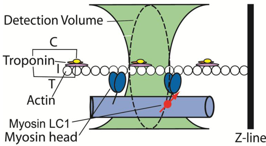
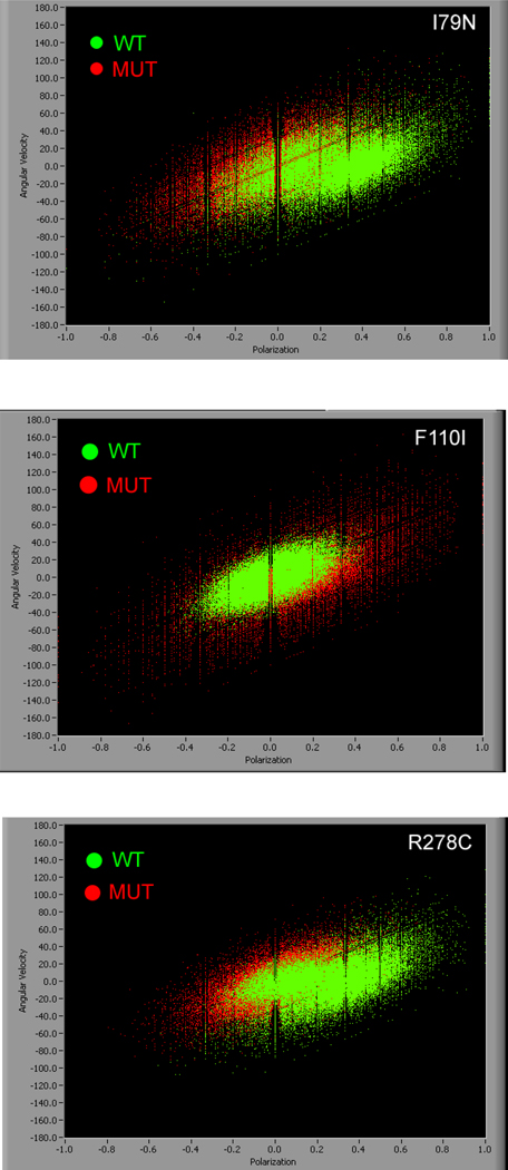
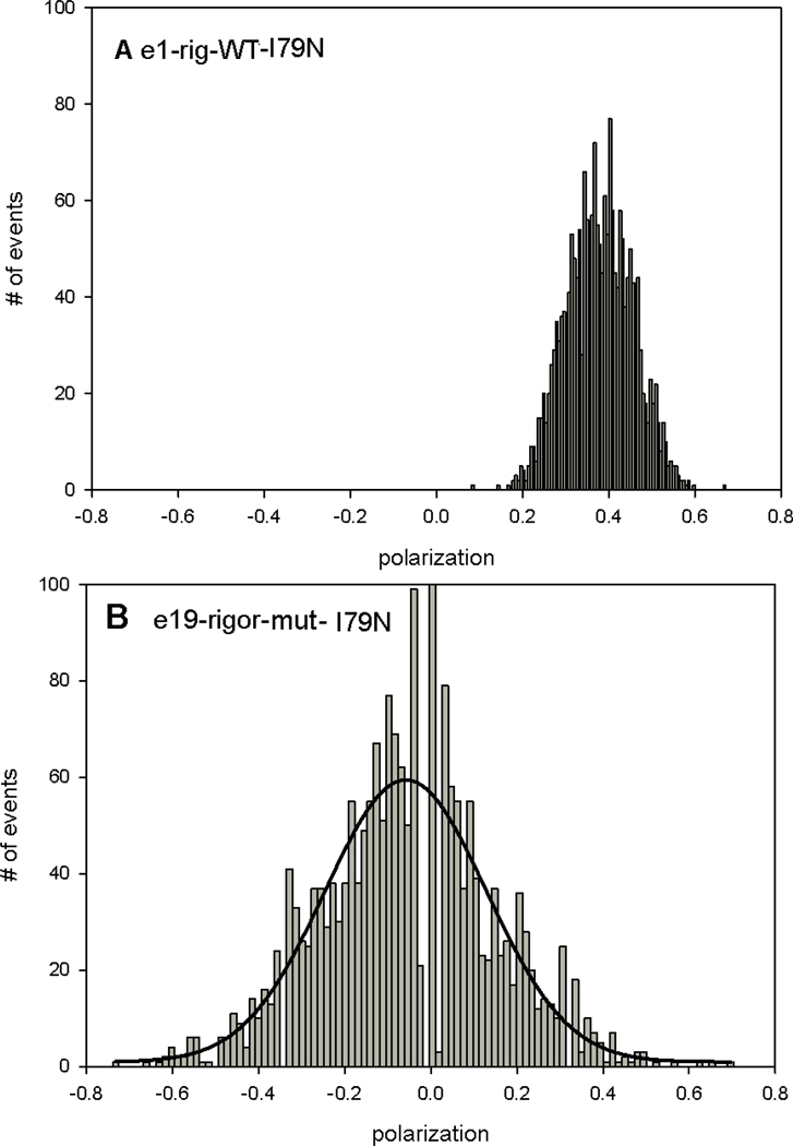
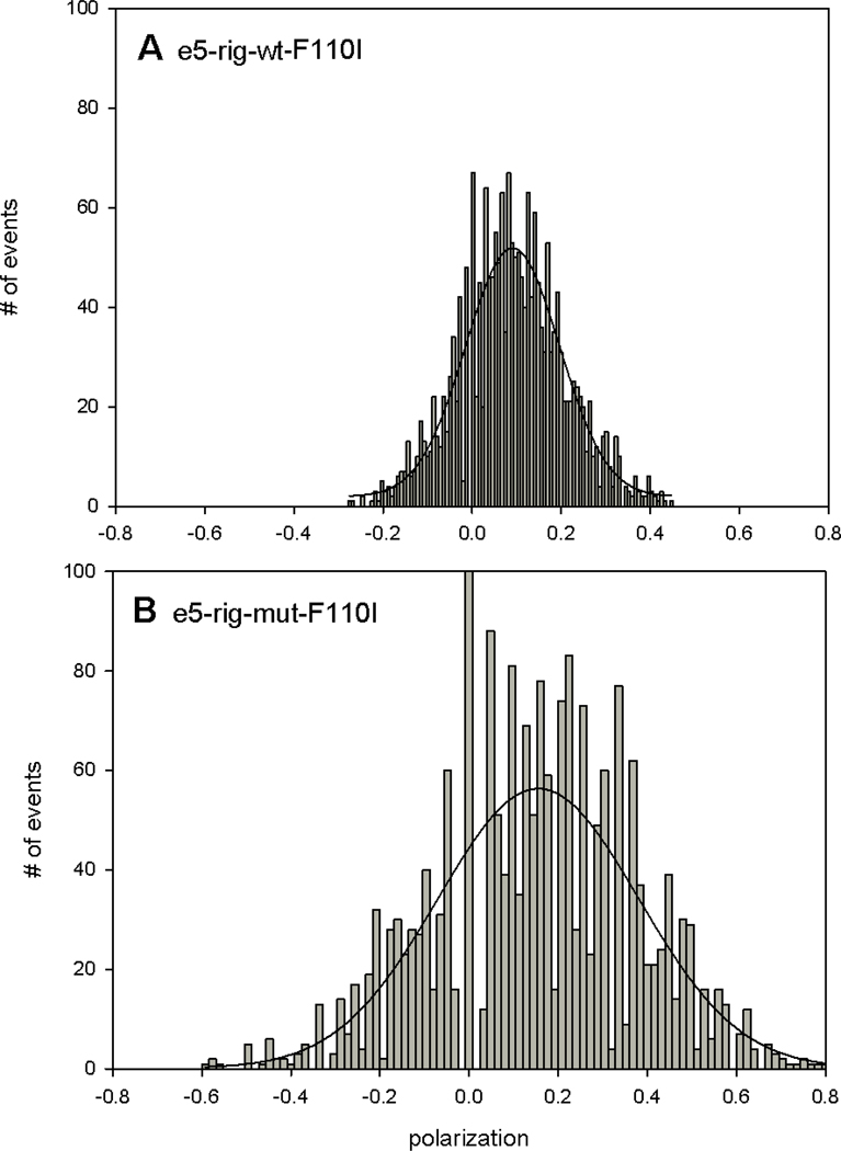
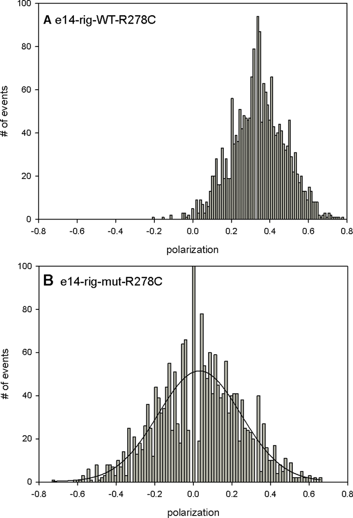
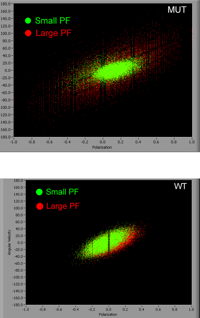
Distribution of orientations of myosin was examined in ex-vivo myofibrils from hearts of transgenic (Tg) mice expressing Familial Hypertrophic Cardiomyopathy (FHC) troponin T (TnT) mutations I79N, F110I and R278C. Humans are heterozygous for sarcomeric FHC mutations and so hypertrophic myocardium contains a mixture of the wild-type (WT) and mutated (MUT) TnT. If mutations are expressed at a low level there may not be a significant change in the global properties of heart muscle. In contrast, measurements from a few molecules avoid averaging inherent in the global measurements. It is thus important to examine the properties of only a few molecules of muscle. To this end, the lever arm of one out of every 60,000 myosin molecules was labeled with a fluorescent dye and a small volume within the A-band (~1 fL) was observed by confocal microscopy. This volume contained on average 5 fluorescent myosin molecules. The lever arm assumes different orientations reflecting different stages of acto-myosin enzymatic cycle. We measured the distribution of these orientations by recording polarization of fluorescent light emitted by myosin-bound fluorophore during rigor and contraction. The distribution of orientations of rigor WT and MUT myofibrils was significantly different. There was a large difference in the width and of skewness and kurtosis of rigor distributions. These findings suggest that the hypertrophic phenotype associated with the TnT mutations can be characterized by a significant increase in disorder of rigor cross-bridges.
我们检测了在表达家族性肥厚型心肌病(FHC)肌钙蛋白 T(TnT)突变 I79N、F110I 和 R278C 的转基因(Tg)小鼠的离体肌原纤维中肌球蛋白取向的分布。人类杂合子存在肌节 FHC 突变,因此肥厚性心肌包含野生型(WT)和突变型(MUT)TnT 的混合物。如果突变以低水平表达,则心肌的整体性质可能不会发生显著变化。相比之下,来自少数分子的测量避免了全局测量中固有的平均化。因此,检查少数几个肌球蛋白分子的特性很重要。为此,每 60000 个肌球蛋白分子中就有一个的臂突用荧光染料标记,并通过共聚焦显微镜观察 A 带内的一小体积(约 1 fL)。该体积平均包含 5 个荧光肌球蛋白分子。臂突呈现出不同的取向,反映了肌球蛋白-肌动蛋白酶循环的不同阶段。我们通过记录肌球蛋白结合荧光团在僵硬和收缩过程中发出的荧光光的偏振来测量这些取向的分布。僵硬 WT 和 MUT 肌原纤维的取向分布有显著差异。僵硬分布的宽度、偏度和峰度有很大差异。这些发现表明,与 TnT 突变相关的肥厚表型可以通过僵硬交联桥的无序程度显著增加来表征。