Infectious Diseases Program, Department of Microbiology, Yong Loo Lin School of Medicine, National University of Singapore, Kent Ridge, Singapore.
Am J Pathol. 2011 Jul;179(1):199-210. doi: 10.1016/j.ajpath.2011.03.013. Epub 2011 May 7.
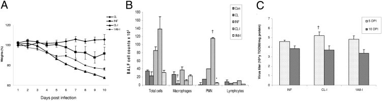
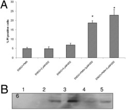
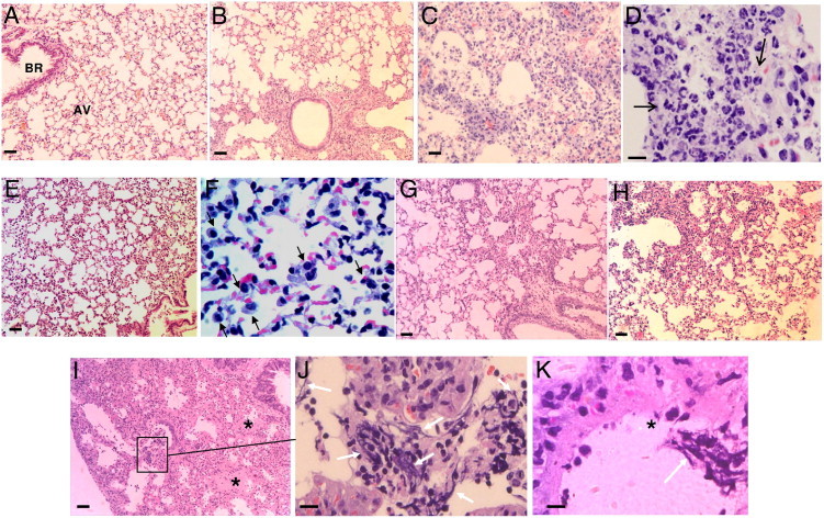
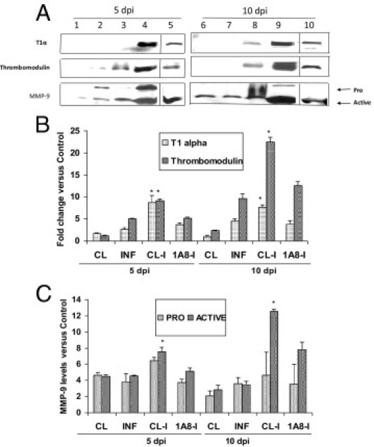
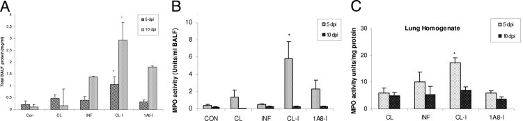
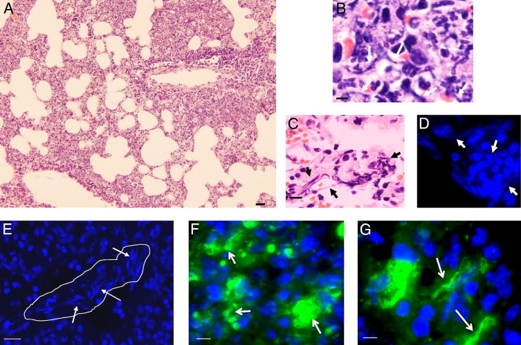
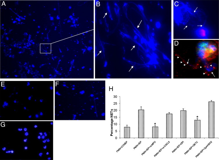
Complications of acute respiratory distress syndrome (ARDS) are common among critically ill patients infected with highly pathogenic influenza viruses. Macrophages and neutrophils constitute the majority of cells recruited into infected lungs, and are associated with immunopathology in influenza pneumonia. We examined pathological manifestations in models of macrophage- or neutrophil-depleted mice challenged with sublethal doses of influenza A virus H1N1 strain PR8. Infected mice depleted of macrophages displayed excessive neutrophilic infiltration, alveolar damage, and increased viral load, later progressing into ARDS-like pathological signs with diffuse alveolar damage, pulmonary edema, hemorrhage, and hypoxemia. In contrast, neutrophil-depleted animals showed mild pathology in lungs. The brochoalveolar lavage fluid of infected macrophage-depleted mice exhibited elevated protein content, T1-α, thrombomodulin, matrix metalloproteinase-9, and myeloperoxidase activities indicating augmented alveolar-capillary damage, compared to neutrophil-depleted animals. We provide evidence for the formation of neutrophil extracellular traps (NETs), entangled with alveoli in areas of tissue injury, suggesting their potential link with lung damage. When co-incubated with infected alveolar epithelial cells in vitro, neutrophils from infected lungs strongly induced NETs generation, and augmented endothelial damage. NETs induction was abrogated by anti-myeloperoxidase antibody and an inhibitor of superoxide dismutase, thus implying that NETs generation is induced by redox enzymes in influenza pneumonia. These findings support the pathogenic effects of excessive neutrophils in acute lung injury of influenza pneumonia by instigating alveolar-capillary damage.
急性呼吸窘迫综合征(ARDS)并发症在感染高致病性流感病毒的重症患者中很常见。巨噬细胞和中性粒细胞构成了感染肺部募集的大多数细胞,与流感肺炎的免疫病理学有关。我们检查了用亚致死剂量的流感 A 病毒 H1N1 株 PR8 感染的巨噬细胞或中性粒细胞耗竭小鼠模型中的病理表现。感染后巨噬细胞耗竭的小鼠显示过度的中性粒细胞浸润、肺泡损伤和病毒载量增加,随后进展为类似于 ARDS 的病理特征,包括弥漫性肺泡损伤、肺水肿、出血和低氧血症。相比之下,中性粒细胞耗竭的动物肺部病变较轻。感染的巨噬细胞耗竭小鼠的支气管肺泡灌洗液中含有较高的蛋白质含量、T1-α、血栓调节蛋白、基质金属蛋白酶-9 和髓过氧化物酶活性,表明肺泡毛细血管损伤加重,与中性粒细胞耗竭动物相比。我们提供了形成中性粒细胞胞外陷阱(NETs)的证据,这些 NETs 在组织损伤区域与肺泡缠绕在一起,表明它们可能与肺损伤有关。当与体外感染的肺泡上皮细胞共孵育时,来自感染肺部的中性粒细胞强烈诱导 NETs 的产生,并增加内皮细胞损伤。NETs 的诱导被抗髓过氧化物酶抗体和超氧化物歧化酶抑制剂所阻断,因此表明 NETs 的产生是由流感肺炎中的氧化还原酶诱导的。这些发现支持了中性粒细胞在流感肺炎急性肺损伤中的致病作用,通过引发肺泡毛细血管损伤。