• 文献检索
  • 文档翻译
  • 深度研究
  • 学术资讯
  • Suppr Zotero 插件Zotero 插件
  • 邀请有礼
  • 套餐&价格
  • 历史记录
应用&插件
Suppr Zotero 插件Zotero 插件浏览器插件Mac 客户端Windows 客户端微信小程序
定价
高级版会员购买积分包购买API积分包
服务
文献检索文档翻译深度研究API 文档MCP 服务
关于我们
关于 Suppr公司介绍联系我们用户协议隐私条款
关注我们

Suppr 超能文献

核心技术专利:CN118964589B侵权必究
粤ICP备2023148730 号-1Suppr @ 2026

文献检索

告别复杂PubMed语法,用中文像聊天一样搜索,搜遍4000万医学文献。AI智能推荐,让科研检索更轻松。

立即免费搜索

文件翻译

保留排版,准确专业,支持PDF/Word/PPT等文件格式,支持 12+语言互译。

免费翻译文档

深度研究

AI帮你快速写综述,25分钟生成高质量综述,智能提取关键信息,辅助科研写作。

立即免费体验

在人体旁路和兔去内皮化模型中捕获循环内皮祖细胞并加速生物工程支架的再内皮化。

Capture of circulatory endothelial progenitor cells and accelerated re-endothelialization of a bio-engineered stent in human ex vivo shunt and rabbit denudation model.

机构信息

Molecular Cardiology Laboratory, Ee2389a, Experimental Cardiology, Thoraxcenter, Erasmus University Medical Center, 's-Gravendijkwal 230, 3015 GE Rotterdam, The Netherlands.

出版信息

Eur Heart J. 2012 Jan;33(1):120-8. doi: 10.1093/eurheartj/ehr196. Epub 2011 Jul 6.

DOI:10.1093/eurheartj/ehr196
PMID:21733913
原文链接:https://pmc.ncbi.nlm.nih.gov/articles/PMC3249218/
Abstract

AIMS

The Genous™ Bio-engineered R™ stent (GS) aims to promote vascular healing by capture of circulatory endothelial progenitor cells (EPCs) to the surface of the stent struts, resulting in accelerated re-endothelialization. Here, we assessed the function of the GS in comparison to bare-metal stent (BMS), when exposed to the human and animal circulation.

METHODS AND RESULTS

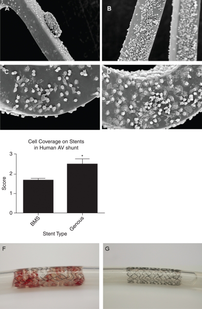
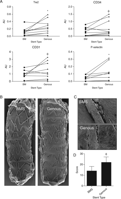
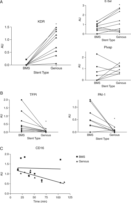
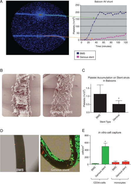
First, 15 patients undergoing coronary angiography received an extracorporeal femoral arteriovenous (AV) shunt containing BMS and GS. Macroscopical mural thrombi were observed in BMS, whereas GS remained visibly clean. Confocal and scanning electron microscopic (SEM) analysis of GS showed an increase in strut coverage. Quantitative polymerase chain reaction (qPCR) analysis of captured cells on the GS demonstrated increased expression of endothelial markers KDR/VEGFR2 and E-selectin, and a decrease in pro-thrombogenic markers tissue factor pathway inhibitor and plasminogen activator inhibitor-1 compared with BMS. Secondly, a similar primate AV shunt model was used to validate these findings and occlusion of BMS was observed, while GS remained patent, as demonstrated by live imaging of indium-labelled platelets. Thirdly, in an in vitro cell-capture assay, GS struts showed increased coverage by EPCs, whereas monocyte coverage remained similar to BMS. Finally, the assessment of re-endothelialization was studied in a rabbit denudation model. Twenty animals received BMS and GS in the aorta and iliac arteries for 7 days. Scanning electron microscopic analysis showed a trend towards increased strut coverage, confirmed by qPCR analysis revealing increased levels of endothelial markers (Tie2, CD34, PCD31, and P-selectin) in GS.

CONCLUSION

In this proof-of-concept study, we have demonstrated that the bio-engineered EPC-capture stent, Genous™ R™ stent, is effective in EPC capture, resulting in accelerated re-endothelialization and reduced thrombogenicity.

摘要

目的

Genous™ 生物工程 R™ 支架(GS)旨在通过捕获循环内皮祖细胞(EPC)到支架支柱的表面来促进血管愈合,从而加速再内皮化。在这里,我们将 GS 与裸金属支架(BMS)进行了比较,评估了它们在暴露于人体和动物循环时的功能。

方法和结果

首先,15 名接受冠状动脉造影的患者接受了包含 BMS 和 GS 的体外股动静脉(AV)分流术。在 BMS 中观察到宏观壁血栓,而 GS 仍然明显干净。GS 的共聚焦和扫描电子显微镜(SEM)分析显示支架覆盖率增加。对 GS 上捕获细胞的定量聚合酶链反应(qPCR)分析表明,与 BMS 相比,内皮标记物 KDR/VEGFR2 和 E-选择素的表达增加,而促血栓形成标记物组织因子途径抑制剂和纤溶酶原激活物抑制剂-1 的表达减少。其次,使用类似的灵长类动物 AV 分流模型验证了这些发现,并观察到 BMS 闭塞,而 GS 保持通畅,这通过铟标记血小板的活体成像得到证实。第三,在体外细胞捕获实验中,GS 支架显示出 EPC 覆盖的增加,而单核细胞覆盖与 BMS 相似。最后,在兔剥脱模型中研究了再内皮化的评估。20 只动物在主动脉和髂动脉中接受 BMS 和 GS 治疗 7 天。扫描电子显微镜分析显示出增加的支架覆盖率趋势,qPCR 分析证实了这一点,结果显示内皮标记物(Tie2、CD34、PCD31 和 P-选择素)水平升高。

结论

在这项概念验证研究中,我们已经证明了生物工程 EPC 捕获支架 Genous™ R™ 支架在 EPC 捕获方面是有效的,从而加速再内皮化并降低血栓形成性。

https://cdn.ncbi.nlm.nih.gov/pmc/blobs/0c3f/3249218/011b90bd60c2/ehr19604.jpg
https://cdn.ncbi.nlm.nih.gov/pmc/blobs/0c3f/3249218/feb1c126c63a/ehr19601.jpg
https://cdn.ncbi.nlm.nih.gov/pmc/blobs/0c3f/3249218/63b50851ef93/ehr19602.jpg
https://cdn.ncbi.nlm.nih.gov/pmc/blobs/0c3f/3249218/5b8c4717d8b9/ehr19603.jpg
https://cdn.ncbi.nlm.nih.gov/pmc/blobs/0c3f/3249218/011b90bd60c2/ehr19604.jpg

相似文献

1
Capture of circulatory endothelial progenitor cells and accelerated re-endothelialization of a bio-engineered stent in human ex vivo shunt and rabbit denudation model.在人体旁路和兔去内皮化模型中捕获循环内皮祖细胞并加速生物工程支架的再内皮化。
Eur Heart J. 2012 Jan;33(1):120-8. doi: 10.1093/eurheartj/ehr196. Epub 2011 Jul 6.
2
The Genous™ endothelial progenitor cell capture stent accelerates stent re-endothelialization but does not affect intimal hyperplasia in porcine coronary arteries.Genous™ 内皮祖细胞捕获支架可加速支架再内皮化,但不影响猪冠状动脉内膜增生。
Catheter Cardiovasc Interv. 2012 Feb 1;79(2):231-42. doi: 10.1002/ccd.22928. Epub 2011 Aug 10.
3
Final results of the HEALING IIB trial to evaluate a bio-engineered CD34 antibody coated stent (Genous™Stent) designed to promote vascular healing by capture of circulating endothelial progenitor cells in CAD patients.评估一种生物工程 CD34 抗体涂层支架(Genous™Stent)的 HEALING IIB 试验的最终结果,该支架旨在通过捕获 CAD 患者循环中的内皮祖细胞来促进血管愈合。
Atherosclerosis. 2011 Nov;219(1):245-52. doi: 10.1016/j.atherosclerosis.2011.06.032. Epub 2011 Jun 25.
4
Stent coated with antibody against vascular endothelial-cadherin captures endothelial progenitor cells, accelerates re-endothelialization, and reduces neointimal formation.抗体涂层支架捕获血管内皮钙黏蛋白,促进内皮祖细胞再内皮化,减少新生内膜形成。
Arterioscler Thromb Vasc Biol. 2011 Dec;31(12):2798-805. doi: 10.1161/ATVBAHA.111.226134. Epub 2011 Oct 20.
5
Combination coating of chitosan and anti-CD34 antibody applied on sirolimus-eluting stents can promote endothelialization while reducing neointimal formation.壳聚糖与抗 CD34 抗体联合涂层应用于雷帕霉素洗脱支架可促进内皮化,同时减少新生内膜形成。
BMC Cardiovasc Disord. 2012 Oct 26;12:96. doi: 10.1186/1471-2261-12-96.
6
Anti-CD34 antibodies immobilized on the surface of sirolimus-eluting stents enhance stent endothelialization.抗 CD34 抗体固定在西罗莫司洗脱支架表面可增强支架内皮化。
JACC Cardiovasc Interv. 2010 Jan;3(1):68-75. doi: 10.1016/j.jcin.2009.09.015.
7
Applying the National Institute for Clinical Excellence criteria to patients treated with the Genous™ Bio-engineered R stent™: a sub-study of the e-HEALING (Healthy Endothelial Accelerated Lining Inhibits Neointimal Growth) worldwide registry.将英国国家临床优化研究所的标准应用于接受Genous™生物工程R支架™治疗的患者:e-HEALING(健康内皮加速内膜抑制新生内膜生长)全球注册研究的一项子研究。
Heart Vessels. 2012 Jul;27(4):360-9. doi: 10.1007/s00380-011-0167-8. Epub 2011 Jul 2.
8
A novel drug-eluting stent coated with an integrin-binding cyclic Arg-Gly-Asp peptide inhibits neointimal hyperplasia by recruiting endothelial progenitor cells.一种涂有整合素结合环Arg-Gly-Asp肽的新型药物洗脱支架通过募集内皮祖细胞抑制内膜增生。
J Am Coll Cardiol. 2006 May 2;47(9):1786-95. doi: 10.1016/j.jacc.2005.11.081. Epub 2006 Apr 19.
9
Circulating endothelial progenitor cells are inversely correlated with in-stent restenosis in patients with non-ST-segment elevation acute coronary syndromes treated with EPC-capture stents (JACK-EPC trial).在接受内皮祖细胞捕获支架治疗的非ST段抬高型急性冠状动脉综合征患者中,循环内皮祖细胞与支架内再狭窄呈负相关(JACK-EPC试验)。
Minerva Cardioangiol. 2013 Jun;61(3):301-11.
10
Late endothelial progenitor cell-capture stents with CD146 antibody and nanostructure reduce in-stent restenosis and thrombosis.带有CD146抗体和纳米结构的晚期内皮祖细胞捕获支架可减少支架内再狭窄和血栓形成。
Acta Biomater. 2020 Jul 15;111:91-101. doi: 10.1016/j.actbio.2020.05.011. Epub 2020 May 17.

引用本文的文献

1
Comparison of very early-phase vascular response to the CD34 antibody‑covered sirolimus‑eluting stent versus durable polymer-coated everolimus-eluting stent.CD34抗体包被的西罗莫司洗脱支架与耐用聚合物涂层依维莫司洗脱支架的极早期血管反应比较。
Cardiovasc Interv Ther. 2025 Apr 18. doi: 10.1007/s12928-025-01114-9.
2
Camouflaging endovascular stents with an endothelial coat using CD31 domain 1 and 2 mimetic peptides.使用CD31结构域1和2模拟肽为血管内支架披上内皮外衣。
JVS Vasc Sci. 2024 Jul 23;5:100213. doi: 10.1016/j.jvssci.2024.100213. eCollection 2024.
3
Development of an In Vitro Blood Vessel Model Using Autologous Endothelial Cells Generated from Footprint-Free hiPSCs to Analyze Interactions of the Endothelium with Blood Cell Components and Vascular Implants.

本文引用的文献

1
Clinical results after coronary stenting with the Genous™ Bio-engineered R stent™: 12-month outcomes of the e-HEALING (Healthy Endothelial Accelerated Lining Inhibits Neointimal Growth) worldwide registry.Genous™ 生物工程 R 支架™ 冠状动脉支架置入术后的临床结果:全球 e-HEALING(健康内皮加速内膜抑制新生内膜生长)注册研究的 12 个月结果。
EuroIntervention. 2011 Feb;6(7):819-25. doi: 10.4244/EIJV6I7A141.
2
Development of a novel prohealing stent designed to deliver sirolimus from a biodegradable abluminal matrix.研发一种新型的促愈合支架,旨在从可生物降解的血管外基质递送来曲唑。
Circ Cardiovasc Interv. 2010 Jun 1;3(3):257-66. doi: 10.1161/CIRCINTERVENTIONS.109.919936. Epub 2010 May 4.
3
利用无印迹 hiPSC 生成的自体内皮细胞开发体外血管模型,以分析内皮细胞与血细胞成分和血管植入物的相互作用。
Cells. 2023 Apr 22;12(9):1217. doi: 10.3390/cells12091217.
4
Biofunctionalization of cardiovascular stents to induce endothelialization: Implications for in- stent thrombosis in diabetes.心血管支架的生物功能化以诱导内皮化:对糖尿病患者支架内血栓形成的影响。
Front Pharmacol. 2022 Oct 10;13:982185. doi: 10.3389/fphar.2022.982185. eCollection 2022.
5
Syndecan-4 and stromal cell-derived factor-1 alpha functionalized endovascular scaffold facilitates adhesion, spreading and differentiation of endothelial colony forming cells and functions under flow and shear stress conditions.硫酸乙酰肝素蛋白聚糖-4 和基质细胞衍生因子-1α 功能化的血管内支架促进内皮祖细胞的黏附、铺展和分化,并在流动和切应力条件下发挥作用。
J Biomed Mater Res B Appl Biomater. 2023 Mar;111(3):538-550. doi: 10.1002/jbm.b.35170. Epub 2022 Oct 8.
6
Optimal duration for dual antiplatelet therapy with COMBO dual therapy stent.COMBO双药治疗支架双重抗血小板治疗的最佳持续时间。
J Geriatr Cardiol. 2019 Nov;16(11):840-843. doi: 10.11909/j.issn.1671-5411.2019.11.001.
7
Assessment of the healing process after percutaneous implantation of a cardiovascular device: a systematic review.经皮植入心血管装置后愈合过程的评估:一项系统综述。
Int J Cardiovasc Imaging. 2020 Mar;36(3):385-394. doi: 10.1007/s10554-019-01734-2. Epub 2019 Nov 19.
8
and evaluation of a dextran-graft-polybutylmethacrylate copolymer coated on CoCr metallic stent.以及对涂覆在钴铬金属支架上的葡聚糖接枝聚甲基丙烯酸丁酯共聚物的评估。
Bioimpacts. 2019;9(1):25-36. doi: 10.15171/bi.2019.04. Epub 2018 Oct 2.
9
Upregulation of miR-126-3p promotes human saphenous vein endothelial cell proliferation and prevents vein graft neointimal formation and .miR-126-3p的上调促进人隐静脉内皮细胞增殖并预防静脉移植物新生内膜形成。
Oncotarget. 2017 Nov 3;8(63):106790-106806. doi: 10.18632/oncotarget.22365. eCollection 2017 Dec 5.
10
Biocompatibility of Coronary Stents.冠状动脉支架的生物相容性
Materials (Basel). 2014 Jan 28;7(2):769-786. doi: 10.3390/ma7020769.
Genous endothelial progenitor cell capturing stent vs. the Taxus Liberte stent in patients with de novo coronary lesions with a high-risk of coronary restenosis: a randomized, single-centre, pilot study.
载基因内皮祖细胞捕获支架与 Taxus Liberte 支架治疗冠状动脉初发病变高危再狭窄患者的随机、单中心、初步研究。
Eur Heart J. 2010 May;31(9):1055-64. doi: 10.1093/eurheartj/ehp476. Epub 2009 Nov 18.
4
Accelerated vascular repair following percutaneous coronary intervention by capture of endothelial progenitor cells promotes regression of neointimal growth at long term follow-up: final results of the Healing II trial using an endothelial progenitor cell capturing stent (Genous R stent).经皮冠状动脉介入治疗后通过捕获内皮祖细胞加速血管修复可促进长期随访时新生内膜生长的消退:使用内皮祖细胞捕获支架(Genous R支架)的Healing II试验的最终结果
EuroIntervention. 2007 Nov;3(3):350-8. doi: 10.4244/eijv3i3a64.
5
Circulating endothelial progenitor cells predict angiographic and intravascular ultrasound outcome following percutaneous coronary interventions in the HEALING-II trial: evaluation of an endothelial progenitor cell capturing stent.在HEALING-II试验中,循环内皮祖细胞可预测经皮冠状动脉介入治疗后的血管造影和血管内超声结果:内皮祖细胞捕获支架的评估
EuroIntervention. 2007 May;3(1):67-75.
6
Genous endothelial progenitor cell-capturing stent system: a novel stent technology.吉诺斯内皮祖细胞捕获支架系统:一种新型支架技术。
Expert Rev Med Devices. 2009 Jul;6(4):365-75. doi: 10.1586/erd.09.16.
7
"Message in the platelet"--more than just vestigial mRNA!“血小板中的信息”——不仅仅是残留的信使核糖核酸!
Platelets. 2008 Sep;19(6):395-404. doi: 10.1080/09537100801990582.
8
Use of endothelial progenitor cell capture stent (Genous Bio-Engineered R Stent) during primary percutaneous coronary intervention in acute myocardial infarction: intermediate- to long-term clinical follow-up.急性心肌梗死直接经皮冠状动脉介入治疗期间使用内皮祖细胞捕获支架(Genous生物工程R支架):中长期临床随访
Am Heart J. 2008 Jan;155(1):128-32. doi: 10.1016/j.ahj.2007.08.031. Epub 2007 Nov 26.
9
Cell therapy for acute myocardial infarction.急性心肌梗死的细胞治疗
Med Clin North Am. 2007 Jul;91(4):769-85; xiii. doi: 10.1016/j.mcna.2007.03.003.
10
Gelatinolytic activity in atherosclerotic plaques is highly localized and is associated with both macrophages and smooth muscle cells in vivo.动脉粥样硬化斑块中的明胶酶活性高度局限,且在体内与巨噬细胞和平滑肌细胞均有关联。
Circulation. 2007 Feb 6;115(5):609-16. doi: 10.1161/CIRCULATIONAHA.106.636415. Epub 2007 Jan 22.