Human Genome Research Center, Biosciences Institute, University of São Paulo, São Paulo, Brazil.
Stem Cell Rev Rep. 2012 Jun;8(2):355-62. doi: 10.1007/s12015-011-9297-8.
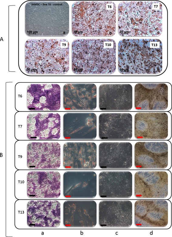
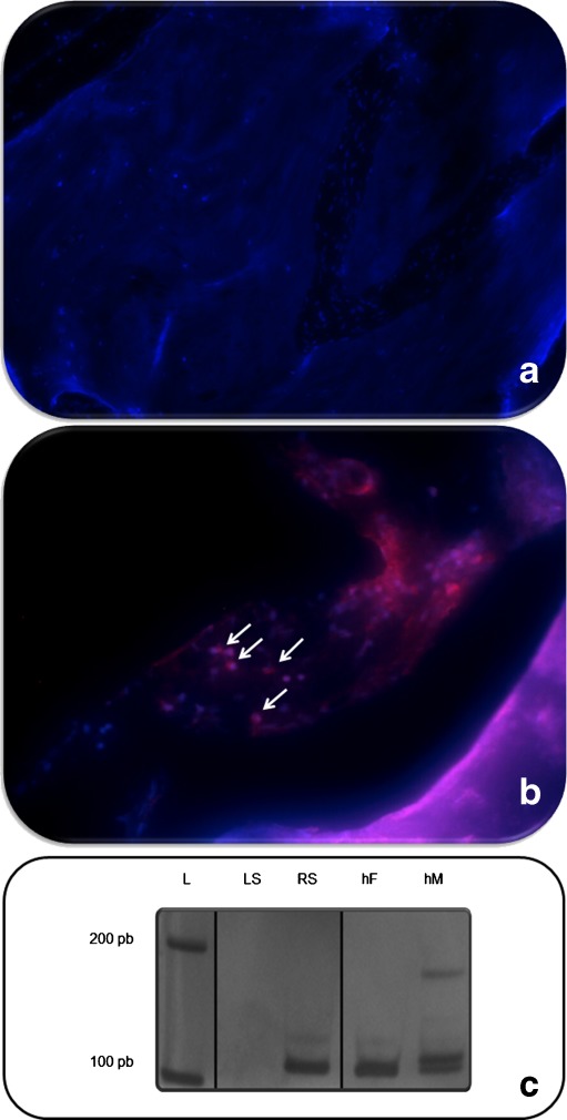
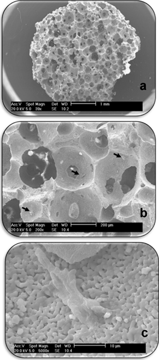
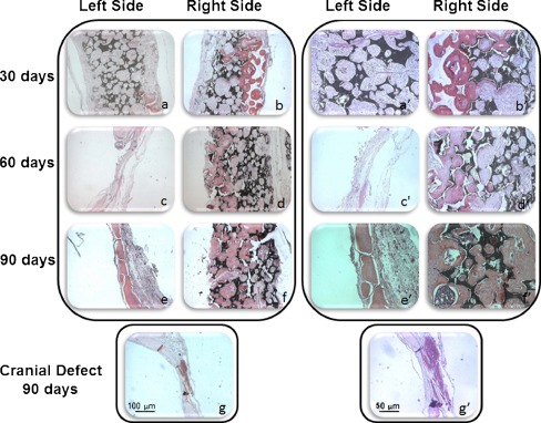
We have recently reported that human fallopian tubes, which are discarded during surgical procedures of women submitted to sterilization or hysterectomies, are a rich source of human fallopian tube mesenchymal stromal cells (htMSCs). It has been previously shown that human mesenchymal stromal cells may be useful in enhancing the speed of bone regeneration. This prompted us to investigate whether htMSCs might be useful for the treatment of osteoporosis or other bone diseases, since they present a pronounced capacity for osteogenic differentiation in vitro. Based on this prior knowledge, our aim was to evaluate, in vivo, the osteogenic capacity of htMSCs to regenerate bone through an already described xenotransplantation model: nonimmunosuppressed (NIS) rats with cranial defects. htMSCs were obtained from five 30-50 years old healthy women and characterized by flow cytometry and for their multipotenciality in vitro capacity (osteogenic, chondrogenic and adipogenic differentiations). Two symmetric full-thickness cranial defects on each parietal region of seven NIS rats were performed. The left side (LS) of six animals was covered with CellCeram (Scaffdex)-a bioabsorbable ceramic composite scaffold that contains 60% hydroxyapatite and 40% β-tricalciumphosphate-only, and the right side (RS) with the CellCeram and htMSCs (10(6) cells/scaffold). The animals were euthanized at 30, 60 and 90 days postoperatively and cranial tissue samples were taken for histological analysis. After 90 days we observed neobone formation in both sides. However, in animals euthanized 30 and 60 days after the procedure, a mature bone was observed only on the side with htMSCs. PCR and immunofluorescence analysis confirmed the presence of human DNA and thus that human cells were not rejected, which further supports the imunomodulatory property of htMSCs. In conclusion, htMSCs can be used successfully to enhance bone regeneration in vivo, opening a new field for future treatments of osteoporosis and bone reconstruction.
我们最近报道称,在接受绝育或子宫切除术的女性的手术过程中丢弃的人类输卵管是人类输卵管间充质基质细胞(htMSCs)的丰富来源。先前已经表明,人类间充质基质细胞可能有助于加速骨再生。这促使我们研究 htMSCs 是否可用于治疗骨质疏松症或其他骨骼疾病,因为它们在体外具有明显的成骨分化能力。基于这一先前的知识,我们的目的是通过已经描述的异种移植模型:无免疫抑制(NIS)大鼠颅骨缺损,体内评估 htMSCs 再生骨的成骨能力。htMSCs 从 5 名 30-50 岁的健康女性中获得,并通过流式细胞术和体外多能性进行特征鉴定(成骨、软骨和成脂分化)。在 7 只 NIS 大鼠的每侧顶骨区域进行两个对称的全厚颅骨缺损。6 只动物的左侧(LS)用 CellCeram(Scaffdex)-一种生物可吸收陶瓷复合支架覆盖,该支架仅包含 60%的羟基磷灰石和 40%的β-磷酸三钙,右侧(RS)用 CellCeram 和 htMSCs(10(6)细胞/支架)覆盖。动物在手术后 30、60 和 90 天被安乐死,取颅骨组织样本进行组织学分析。90 天后,我们在两侧均观察到新骨形成。然而,在手术后 30 和 60 天安乐死的动物中,仅在有 htMSCs 的一侧观察到成熟骨。PCR 和免疫荧光分析证实了人 DNA 的存在,因此表明人类细胞未被排斥,这进一步支持了 htMSCs 的免疫调节特性。总之,htMSCs 可成功用于增强体内骨再生,为骨质疏松症和骨重建的未来治疗开辟了新领域。