Department of Vascular Surgery, Cleveland Clinic Lerner College of Medicine-CWRU, Cleveland, Ohio 44195, USA.
J Surg Res. 2012 Jul;176(1):321-8. doi: 10.1016/j.jss.2011.05.041. Epub 2011 Jun 22.
Signal transducers and activators of transcription (STAT) proteins are transcription factors that, when activated by phosphorylation, regulate gene expression and cellular activity. The aim of this study was to evaluate the local and systemic expression and activation of STAT proteins associated with abdominal aortic aneurysms (AAA).
Expression and activation of STAT proteins were assessed in aortic wall samples obtained from patients undergoing repair of AAA (n = 9) and from non-aneurysmal (NA) donors (n = 17). Aortic samples were evaluated for mRNA and protein expression for STAT1, 2, 3, 4, 5a, and 5b using RT-PCR and immunoblot (WB) assays and normalized to ß-actin (expressed as arbitrary units). STAT activation was assessed with WB assays using phosphorylated (p)-STAT-specific antibodies. Alterations in STAT activation were calculated by normalizing pSTAT proteins to corresponding total STAT levels. Immunohistochemistry was performed on AAA and NA samples using the total and pSTAT antibodies. Systemic alterations in STAT activation were assessed by evaluating circulating leukocytes for the presence of pSTAT from patients with AAA (AAA, n = 8), repaired aneurysm (RA, n = 8), or age/gender matched controls with no AAA (CT, n = 8). Flow cytometry was performed to assess for circulating levels of STAT1 (pY701), STAT3 (pY705), and STAT5a (pY694) in monocytes, granulocytes, and lymphocytes. Assessments were made at baseline and in response to in vitro stimulation with IFN-γ (50 ng/mL) or IL-6 (100 ng/mL). Results were analyzed using Student's t-test and are expressed as mean ± SEM.
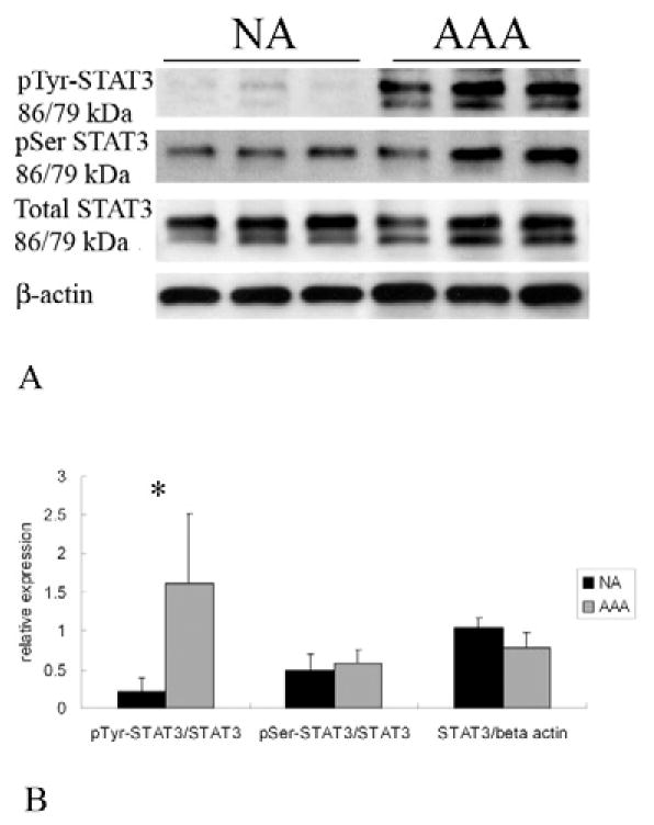
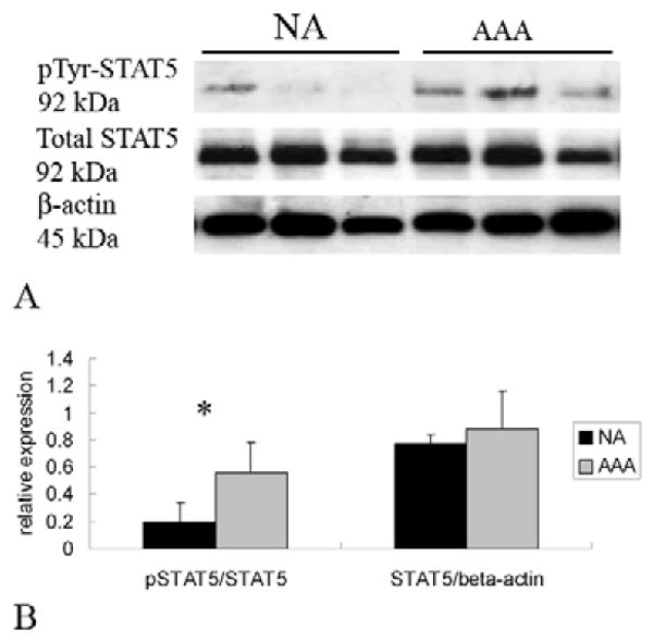
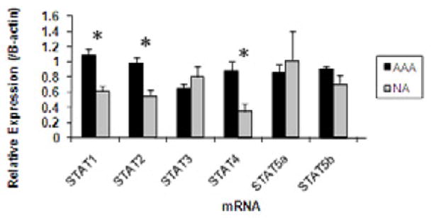
In AAA tissue compared with NA, STAT-1 (1.08 ± 0.09 versus 0.62 ± 0.07), -2 (0.98 ± 0.07 versus 0.55 ± 0.08), and -4 (0.89 ± 0.12 versus 0.35 ± 0.11) mRNA levels were elevated (P < 0.01, all). Corresponding increases in STAT protein were only observed for STAT1 (2.77 ± 0.93 versus 0.93 ± 0.08, P < 0.05). Increases in activation were observed in AAA compared with NA in pSTAT2 (0.77 ± 0.1 versus 0.1 ± 0.02, P < 0.01), pSTAT3 (1.6 ± 0.3 versus 0.2 ± 0.06, P < 0.02) and pSTAT5 (0.57 ± 0.03 versus 0.2 ± 0.03, P < 0.05) levels. Phosphorylated STAT1, 2, 3, and 5 were observed in inflammatory cells invading the AAA adventitia. In addition, STAT3 was observed in the media of AAA and NA, but pSTAT3 was only observed in the media of AAA. There were no differences in baseline levels of pSTAT-positive circulating leukocytes. IFN-γ stimulation decreased STAT-5a (pY694)-positive CT lymphocytes to 40% ± 13% of baseline, but had no effect on AAA or RA lymphocytes (116% ± 35%, 102% ± 19%, respectively; P = 0.01). STAT-5a (pY694)-positive CT granulocytes also decreased to 62% ± 18% of baseline compared with AAA or RA granulocytes (122% ± 25%, 126% ± 17%, respectively; P = 0.01). Alterations in STAT1 (pY701) and STAT3 (pY705) were not observed in leukocytes following cytokine stimulation.
STAT proteins are important regulators of transcriptional activity and have been linked to cardiovascular disease. The present data suggest that altered levels of phosphorylated STATs are associated with AAA. Understanding their role may provide further insight into the mechanisms of AAA formation and allow for the development of medical treatment options.
信号转导和转录激活因子(STAT)蛋白是转录因子,当被磷酸化激活时,可调节基因表达和细胞活性。本研究旨在评估与腹主动脉瘤(AAA)相关的 STAT 蛋白的局部和全身表达和激活。
使用 RT-PCR 和免疫印迹(WB)测定评估从接受 AAA 修复(n = 9)和非动脉瘤(NA)供体(n = 17)获得的主动脉壁样本中 STAT 蛋白的表达和激活。使用磷酸化(p)-STAT 特异性抗体的 WB 测定评估 STAT1、2、3、4、5a 和 5b 的 mRNA 和蛋白表达,并相对于β-肌动蛋白(表示为任意单位)进行归一化。通过将 pSTAT 蛋白与相应的总 STAT 水平进行归一化来评估 STAT 激活的变化。使用总和 pSTAT 抗体对 AAA 和 NA 样本进行免疫组织化学染色。通过评估来自 AAA(AAA,n = 8)、修复的动脉瘤(RA,n = 8)或无 AAA 的年龄/性别匹配对照(CT,n = 8)患者的循环白细胞中 pSTAT 的存在来评估全身性 STAT 激活的变化。通过流式细胞术评估单核细胞、粒细胞和淋巴细胞中 STAT1(pY701)、STAT3(pY705)和 STAT5a(pY694)的循环水平。在基础状态下和体外用 IFN-γ(50ng/mL)或 IL-6(100ng/mL)刺激后进行评估。使用 Student's t 检验进行分析,结果表示为平均值 ± SEM。
与 NA 相比,AAA 组织中的 STAT-1(1.08 ± 0.09 与 0.62 ± 0.07)、-2(0.98 ± 0.07 与 0.55 ± 0.08)和-4(0.89 ± 0.12 与 0.35 ± 0.11)mRNA 水平升高(均 P < 0.01)。仅观察到 STAT1 蛋白相应增加(2.77 ± 0.93 与 0.93 ± 0.08,P < 0.05)。与 NA 相比,AAA 中观察到 pSTAT2(0.77 ± 0.1 与 0.1 ± 0.02,P < 0.01)、pSTAT3(1.6 ± 0.3 与 0.2 ± 0.06,P < 0.02)和 pSTAT5(0.57 ± 0.03 与 0.2 ± 0.03,P < 0.05)水平升高。在 AAA 的炎症细胞中观察到磷酸化 STAT1、2、3 和 5。此外,在 AAA 和 NA 的中膜中观察到 STAT3,但仅在 AAA 的中膜中观察到 pSTAT3。循环白细胞中 pSTAT 阳性细胞的基线水平没有差异。IFN-γ 刺激将 CT 淋巴细胞中 pSTAT-5a(pY694)阳性细胞减少到基线的 40% ± 13%,但对 AAA 或 RA 淋巴细胞无影响(分别为 116% ± 35%、102% ± 19%,P = 0.01)。与 AAA 或 RA 粒细胞相比,CT 粒细胞中 pSTAT-5a(pY694)阳性细胞也减少到基线的 62% ± 18%(分别为 122% ± 25%、126% ± 17%,P = 0.01)。在白细胞中未观察到细胞因子刺激后 STAT1(pY701)和 STAT3(pY705)的改变。
STAT 蛋白是转录活性的重要调节剂,与心血管疾病有关。本研究数据表明,磷酸化 STATs 的水平改变与 AAA 相关。了解它们的作用可能会进一步深入了解 AAA 形成的机制,并为开发医疗治疗方案提供依据。