Department of Immunobiology, Yale University School of Medicine, New Haven, Connecticut 06520, USA.
Nature. 2011 Jul 17;475(7357):514-8. doi: 10.1038/nature10228.
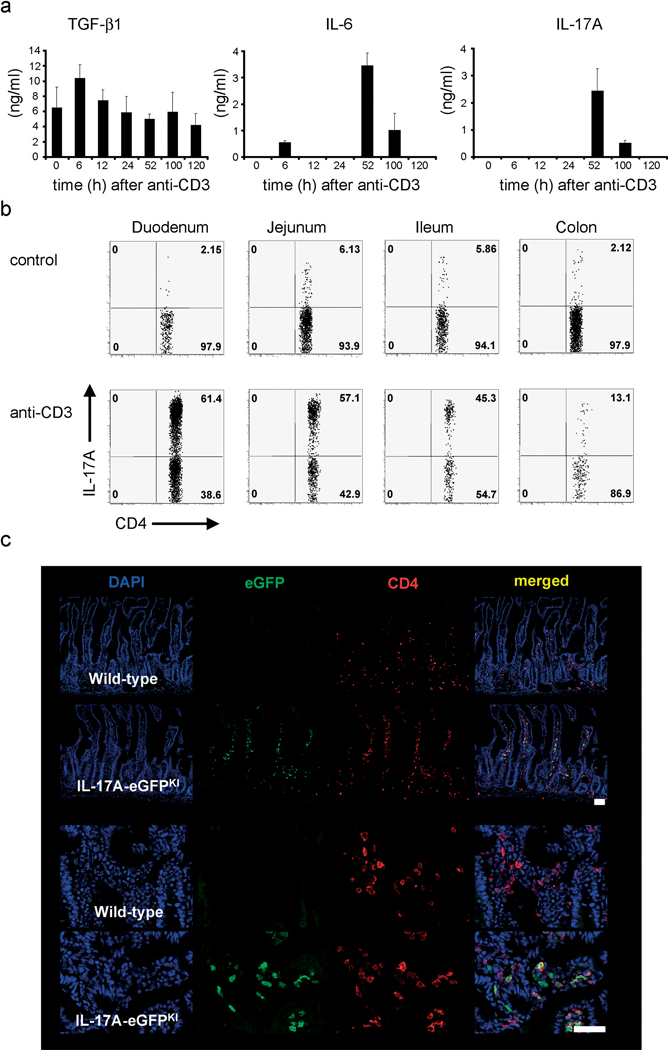
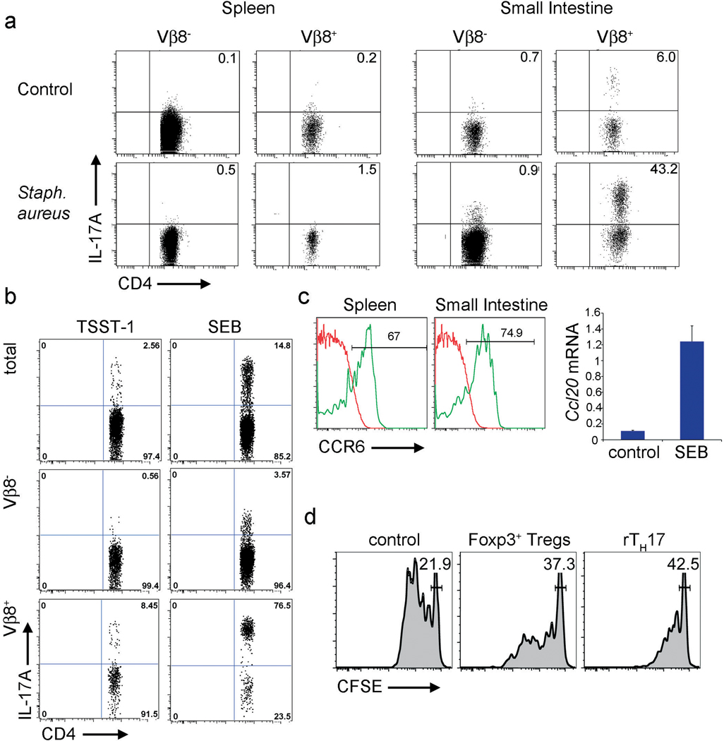
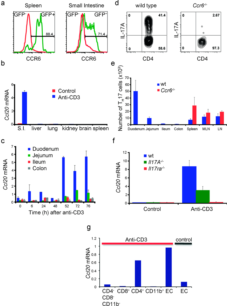
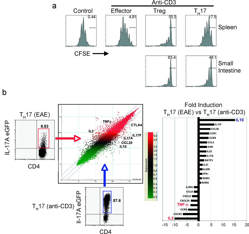
Interleukin (IL)-17-producing T helper cells (T(H)17) are a recently identified CD4(+) T cell subset distinct from T helper type 1 (T(H)1) and T helper type 2 (T(H)2) cells. T(H)17 cells can drive antigen-specific autoimmune diseases and are considered the main population of pathogenic T cells driving experimental autoimmune encephalomyelitis (EAE), the mouse model for multiple sclerosis. The factors that are needed for the generation of T(H)17 cells have been well characterized. However, where and how the immune system controls T(H)17 cells in vivo remains unclear. Here, by using a model of tolerance induced by CD3-specific antibody, a model of sepsis and influenza A viral infection (H1N1), we show that pro-inflammatory T(H)17 cells can be redirected to and controlled in the small intestine. T(H)17-specific IL-17A secretion induced expression of the chemokine CCL20 in the small intestine, facilitating the migration of these cells specifically to the small intestine via the CCR6/CCL20 axis. Moreover, we found that T(H)17 cells are controlled by two different mechanisms in the small intestine: first, they are eliminated via the intestinal lumen; second, pro-inflammatory T(H)17 cells simultaneously acquire a regulatory phenotype with in vitro and in vivo immune-suppressive properties (rT(H)17). These results identify mechanisms limiting T(H)17 cell pathogenicity and implicate the gastrointestinal tract as a site for control of T(H)17 cells.
白细胞介素 (IL)-17 产生的辅助性 T 细胞 (T(H)17) 是最近发现的不同于辅助性 T 细胞 1 (T(H)1) 和辅助性 T 细胞 2 (T(H)2) 的 CD4(+)T 细胞亚群。T(H)17 细胞可驱动抗原特异性自身免疫性疾病,被认为是驱动实验性自身免疫性脑脊髓炎 (EAE) 的主要致病性 T 细胞群体,EAE 是多发性硬化症的小鼠模型。T(H)17 细胞生成所需的因素已得到很好的描述。然而,免疫系统如何在体内控制 T(H)17 细胞仍然不清楚。在这里,我们通过使用 CD3 特异性抗体诱导的耐受模型、脓毒症模型和流感 A 病毒感染 (H1N1) 模型,显示促炎性 T(H)17 细胞可以被重新定向并在小肠中得到控制。T(H)17 细胞特异性的 IL-17A 分泌诱导了小肠中趋化因子 CCL20 的表达,通过 CCR6/CCL20 轴促进这些细胞特异性迁移到小肠。此外,我们发现 T(H)17 细胞在小肠中受到两种不同机制的控制:首先,它们通过肠腔被消除;其次,促炎性 T(H)17 细胞同时获得具有体外和体内免疫抑制特性的调节表型 (rT(H)17)。这些结果确定了限制 T(H)17 细胞致病性的机制,并暗示胃肠道是控制 T(H)17 细胞的部位。