Department of Parasitology, College of Medicine, Inha University, Incheon405-751, Republic of Korea.
Malar J. 2011 Aug 6;10:228. doi: 10.1186/1475-2875-10-228.
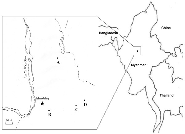
The aim of this study was to investigate the profile of antibodies against several antigens of Plasmodium vivax and Plasmodium falciparum in Mandalay, Myanmar.
Malaria parasites were identified by microscopic examination. To test the antibodies against P. vivax and P. falciparum in sera, an indirect immunofluorescence antibody test (IFAT) was performed using asexual blood parasite antigens. An enzyme-linked immunosorbent assay (ELISA) was performed with circumsporozoite protein (CSP), Pvs25 and Pvs28 recombinant proteins of transmission-blocking vaccine candidates for P. vivax, and liver stage specific antigen-1 and -3 (PfLSA-1, PfLSA-3) for P. falciparum.
Fourteen patients among 112 were found to be infected with P. vivax and 26 with P. falciparum by thick smear examination. Twenty-three patients were found to be infected with P. vivax, 19 with P. falciparum and five with both by thin smear examination. Blood samples were divided into two groups: Group I consisted of patients who were positive for infection by microscopic examination, and Group II consisted of those who showed symptoms, but were negative in microscopic examination. In P. falciparum, IgG against the blood stage antigen in Group I (80.8%) was higher than in Group II (70.0%). In P. vivax, IgG against the blood stage antigen in Group I (53.8%) was higher than in Group II (41.7%). However, the positivity rate of the PvCSP VK210 subtype in Group II (40.0%) was higher than in Group I (23.1%). Similarly for the PvCSP VK247 subtype, Group II (21.7%) was higher than that for Group I (9.6%). A similar pattern was observed in the ELISA using Pvs25 and Pvs28: positive rates of Group II were higher than those for Group I. However, those differences were not shown significant in statistics.
The positive rates for blood stage antigens of P. falciparum were higher in Group I than in Group II, but the positive rates for antigens of other stages (PfLSA-1 and -3) showed opposite results. Similar to P. falciparum, the positive rate of pre-blood stage (CSP VK210 and 247 subtype) and post-blood stage (Pvs25 and 28) antigens of P. vivax were higher in Group II than in Group I. Therefore, sero-diagnosis is not helpful to discriminate between malaria patients and symptomatic individuals during the epidemic season in Myanmar.
本研究旨在调查缅甸曼德勒地区间日疟原虫和恶性疟原虫的几种抗原抗体谱。
通过显微镜检查鉴定疟原虫。采用间接免疫荧光抗体试验(IFAT)检测血清中抗间日疟原虫和恶性疟原虫抗体。采用环子孢子蛋白(CSP)、恶性疟原虫传播阻断候选疫苗的 Pvs25 和 Pvs28 重组蛋白、恶性疟原虫肝期特异抗原-1 和 -3(PfLSA-1、PfLSA-3)的酶联免疫吸附试验(ELISA)检测抗体。
112 例患者中,14 例经厚涂片检查发现感染间日疟原虫,26 例感染恶性疟原虫。23 例患者经薄涂片检查发现感染间日疟原虫,19 例感染恶性疟原虫,5 例同时感染两种疟原虫。血样分为两组:组 I 为镜检阳性的患者,组 II 为有症状但镜检阴性的患者。在恶性疟原虫中,组 I(80.8%)的血期抗原 IgG 高于组 II(70.0%)。在间日疟原虫中,组 I(53.8%)的血期抗原 IgG 高于组 II(41.7%)。然而,组 II(40.0%)的 PvCSP VK210 亚型阳性率高于组 I(23.1%)。同样,对于 PvCSP VK247 亚型,组 II(21.7%)也高于组 I(9.6%)。用 Pvs25 和 Pvs28 进行 ELISA 也观察到类似的模式:组 II 的阳性率高于组 I。然而,这些差异在统计学上并不显著。
间日疟原虫血期抗原在组 I 中的阳性率高于组 II,但其他阶段(PfLSA-1 和 -3)抗原的阳性率则相反。与恶性疟原虫相似,间日疟原虫的前血期(CSP VK210 和 247 亚型)和后血期(Pvs25 和 28)抗原在组 II 中的阳性率高于组 I。因此,在缅甸流行季节,血清诊断无助于区分疟疾患者和有症状的个体。