Department of Molecular and Medical Pharmacology, David Geffen School of Medicine at UCLA, Los Angeles, CA, USA.
EJNMMI Res. 2011 Jul 6;1:8. doi: 10.1186/2191-219X-1-8.
We evaluated the effect of insulin stimulation and dietary changes on myocardial, skeletal muscle and brain [(18)F]-fluorodeoxyglucose (FDG) kinetics and uptake in vivo in intact mice.
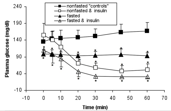
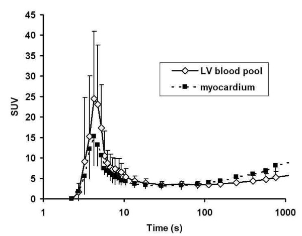
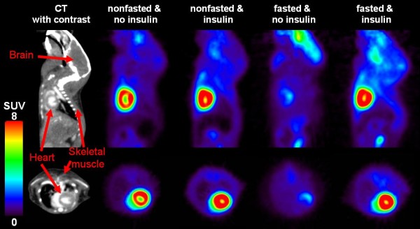
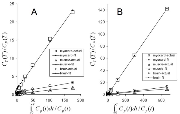
Mice were anesthetized with isoflurane and imaged under different conditions: non-fasted (n = 7; "controls"), non-fasted with insulin (2 IU/kg body weight) injected subcutaneously immediately prior to FDG (n = 6), fasted (n = 5), and fasted with insulin injection (n = 5). A 60-min small-animal PET with serial blood sampling and kinetic modeling was performed.
We found comparable FDG standardized uptake values (SUVs) in myocardium in the non-fasted controls and non-fasted-insulin injected group (SUV 45-60 min, 9.58 ± 1.62 vs. 9.98 ± 2.44; p = 0.74), a lower myocardial SUV was noted in the fasted group (3.48 ± 1.73; p < 0.001). In contrast, the FDG uptake rate constant (K(i)) for myocardium increased significantly by 47% in non-fasted mice by insulin (13.4 ± 3.9 ml/min/100 g vs. 19.8 ± 3.3 ml/min/100 g; p = 0.030); in fasted mice, a lower myocardial K(i) as compared to controls was observed (3.3 ± 1.9 ml/min/100 g; p < 0.001). Skeletal muscle SUVs and K(i) values were increased by insulin independent of dietary state, whereas in the brain, those parameters were not influenced by fasting or administration of insulin. Fasting led to a reduction in glucose metabolic rate in the myocardium (19.41 ± 5.39 vs. 3.26 ± 1.97 mg/min/100 g; p < 0.001), the skeletal muscle (1.06 ± 0.34 vs. 0.34 ± 0.08 mg/min/100 g; p = 0.001) but not the brain (3.21 ± 0.53 vs. 2.85 ±0.25 mg/min/100 g; p = 0.19).
Changes in organ SUVs, uptake rate constants and metabolic rates induced by fasting and insulin administration as observed in intact mice by small-animal PET imaging are consistent with those observed in isolated heart/muscle preparations and, more importantly, in vivo studies in larger animals and in humans. When assessing the effect of insulin on the myocardial glucose metabolism of non-fasted mice, it is not sufficient to just calculate the SUV - dynamic imaging with kinetic modeling is necessary.
我们评估了胰岛素刺激和饮食变化对完整小鼠体内心肌、骨骼肌和脑 [(18)F]-氟脱氧葡萄糖(FDG)动力学和摄取的影响。
小鼠用异氟烷麻醉,并在不同条件下进行成像:未禁食(n = 7;“对照”)、未禁食并在 FDG 前立即皮下注射胰岛素(2 IU/kg 体重)(n = 6)、禁食(n = 5)和禁食并注射胰岛素(n = 5)。进行了 60 分钟小动物 PET 扫描,同时进行了连续的血液采样和动力学建模。
我们发现未禁食对照组和皮下注射胰岛素组的心肌 FDG 标准化摄取值(SUV)相似(SUV 45-60 分钟,9.58 ± 1.62 比 9.98 ± 2.44;p = 0.74),禁食组的心肌 SUV 较低(3.48 ± 1.73;p < 0.001)。相比之下,胰岛素可使未禁食小鼠的心肌 FDG 摄取率常数(K(i))显著增加 47%(13.4 ± 3.9 ml/min/100 g 比 19.8 ± 3.3 ml/min/100 g;p = 0.030);在禁食小鼠中,与对照组相比,心肌 K(i)较低(3.3 ± 1.9 ml/min/100 g;p < 0.001)。胰岛素可使骨骼肌 SUV 和 K(i)值增加,与饮食状态无关,而在大脑中,这些参数不受禁食或胰岛素给药的影响。禁食导致心肌(19.41 ± 5.39 比 3.26 ± 1.97 mg/min/100 g;p < 0.001)、骨骼肌(1.06 ± 0.34 比 0.34 ± 0.08 mg/min/100 g;p = 0.001)但不是大脑(3.21 ± 0.53 比 2.85 ±0.25 mg/min/100 g;p = 0.19)的葡萄糖代谢率降低。
在完整小鼠中通过小动物 PET 成像观察到的禁食和胰岛素给药引起的器官 SUV、摄取率常数和代谢率的变化与在分离的心脏/肌肉制剂中观察到的变化一致,更重要的是,与较大动物和人体中的体内研究一致。在评估非禁食小鼠胰岛素对心肌葡萄糖代谢的影响时,仅仅计算 SUV 是不够的——需要进行动态成像的动力学建模。