Department of Urology, AMC University Hospital, Meibergdreef 9, 1105 AZ Amsterdam, The Netherlands.
World J Urol. 2011 Oct;29(5):581-7. doi: 10.1007/s00345-011-0747-3. Epub 2011 Aug 17.
The purposes of this paper were to present the current status of contrast-enhanced transrectal ultrasound imaging and to discuss the latest achievements and techniques now under preclinical testing.
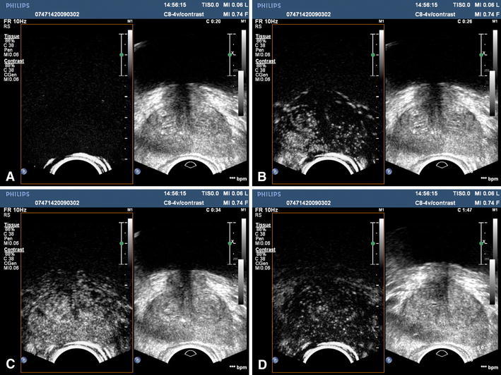
Although grayscale transrectal ultrasound is the standard method for prostate imaging, it lacks accuracy in the detection and localization of prostate cancer. With the introduction of contrast-enhanced ultrasound (CEUS), perfusion imaging of the microvascularization became available. By this, cancer-induced neovascularisation can be visualized with the potential to improve ultrasound imaging for prostate cancer detection and localization significantly. For example, several studies have shown that CEUS-guided biopsies have the same or higher PCa detection rate compared with systematic biopsies with less biopsies needed.
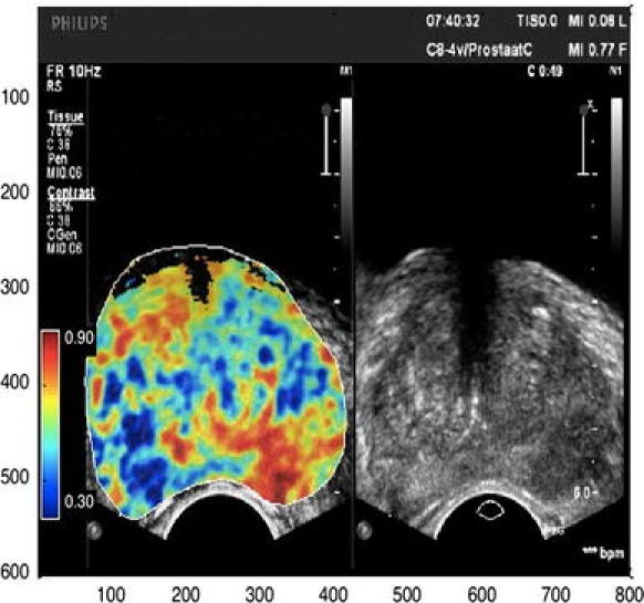
This paper describes the current status of CEUS and discusses novel quantification techniques that can improve the accuracy even further. Furthermore, quantification might decrease the user-dependency, opening the door to use in the routine clinical environment. A new generation of targeted microbubbles is now under pre-clinical testing and showed avidly binding to VEGFR-2, a receptor up-regulated in prostate cancer due to angiogenesis. The first publications regarding a targeted microbubble ready for human use will be discussed.
Ultrasound-assisted drug delivery gives rise to a whole new set of therapeutic options, also for prostate cancer. A major breakthrough in the future can be expected from the clinical use of targeted microbubbles for drug delivery for prostate cancer diagnosis as well as treatment.
本文旨在介绍对比增强经直肠超声成像的现状,并讨论目前处于临床前测试阶段的最新成果和技术。
尽管灰度经直肠超声是前列腺成像的标准方法,但它在前列腺癌的检测和定位方面缺乏准确性。随着对比增强超声(CEUS)的引入,微血管灌注成像成为可能。通过这种方式,可以可视化癌症引起的新生血管化,从而有可能显著提高超声成像在前列腺癌检测和定位方面的性能。例如,多项研究表明,CEUS 引导下的活检与系统活检具有相同或更高的前列腺癌检出率,且所需活检次数更少。
本文描述了 CEUS 的现状,并讨论了可以进一步提高准确性的新型定量技术。此外,定量技术可能会降低对用户的依赖性,为在常规临床环境中使用打开大门。新一代靶向微泡目前正在进行临床前测试,它们可以强烈结合血管内皮生长因子受体 2(VEGFR-2),由于血管生成,前列腺癌中 VEGFR-2 会被上调。本文将讨论关于可用于人体的靶向微泡的首批出版物。
超声辅助药物输送为治疗前列腺癌带来了全新的治疗选择。靶向微泡在前列腺癌诊断和治疗中的临床应用有望成为未来的重大突破。