Department of Psychiatry, University of Wisconsin-Madison, Madison, WI, USA.
Prog Brain Res. 2011;193:17-38. doi: 10.1016/B978-0-444-53839-0.00002-8.
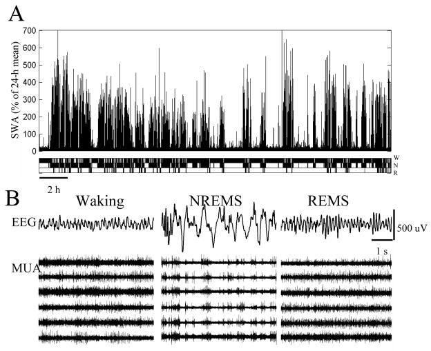
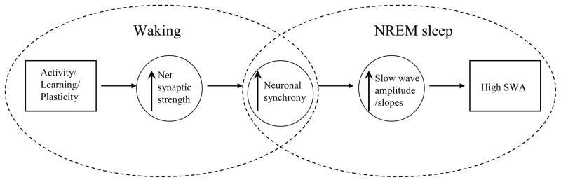
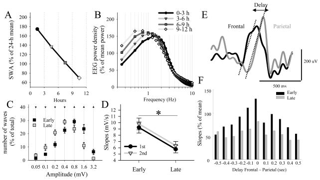
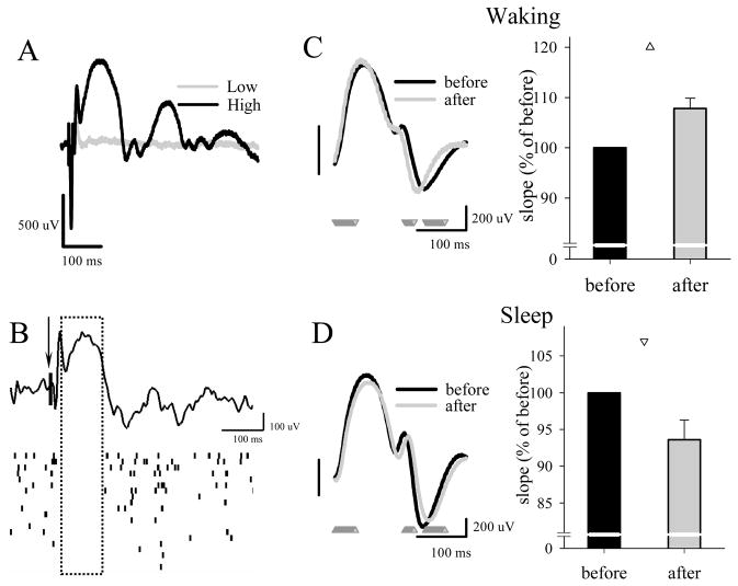
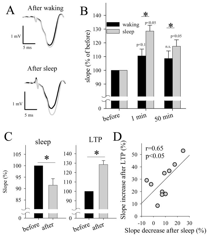
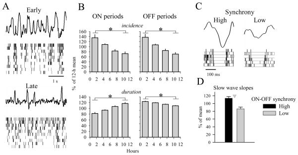
The electrical activity of the brain does not only reflect the current level of arousal, ongoing behavior, or involvement in a specific task but is also influenced by what kind of activity, and how much sleep and waking occurred before. The best marker of sleep-wake history is the electroencephalogram (EEG) spectral power in slow frequencies (slow-wave activity, 0.5-4 Hz, SWA) during sleep, which is high after extended wakefulness and low after consolidated sleep. While sleep homeostasis has been well characterized in various species and experimental paradigms, the specific mechanisms underlying homeostatic changes in brain activity or their functional significance remain poorly understood. However, several recent studies in humans, rats, and computer simulations shed light on the cortical mechanisms underlying sleep regulation. First, it was found that the homeostatic changes in SWA can be fully accounted for by the variations in amplitude and slope of EEG slow waves, which are in turn determined by the efficacy of corticocortical connectivity. Specifically, the slopes of sleep slow waves were steeper in early sleep compared to late sleep. Second, the slope of cortical evoked potentials, which is an established marker of synaptic strength, was steeper after waking, and decreased after sleep. Further, cortical long-term potentiation (LTP) was partially occluded if it was induced after a period of waking, but it could again be fully expressed after sleep. Finally, multiunit activity recordings during sleep revealed that cortical neurons fired more synchronously after waking, and less so after a period of consolidated sleep. The decline of all these electrophysiological measures-the slopes of slow waves and evoked potentials and neuronal synchrony-during sleep correlated with the decline of the traditional marker of sleep homeostasis, EEG SWA. Taken together, these data suggest that homeostatic changes in sleep EEG are the result of altered neuronal firing and synchrony, which in turn arise from changes in functional neuronal connectivity.
大脑的电活动不仅反映了当前的觉醒水平、正在进行的行为或参与特定任务的程度,还受到之前进行了哪种活动以及睡眠和觉醒的时间长短的影响。睡眠-觉醒历史的最佳标志物是睡眠期间的慢频(慢波活动,0.5-4Hz,SWA)脑电图(EEG)谱功率,在长时间觉醒后高,在巩固的睡眠后低。尽管睡眠稳态在各种物种和实验范式中得到了很好的描述,但大脑活动的稳态变化背后的具体机制及其功能意义仍知之甚少。然而,最近在人类、大鼠和计算机模拟中的几项研究揭示了睡眠调节背后的皮层机制。首先,人们发现 SWA 的稳态变化可以完全由 EEG 慢波的幅度和斜率的变化来解释,而这些变化又取决于皮质间连接的效能。具体而言,与晚期睡眠相比,早期睡眠中的睡眠慢波斜率更陡。其次,皮质诱发电位的斜率(突触强度的既定标志物)在醒来后更陡,在睡眠后下降。此外,如果在觉醒后一段时间内诱导皮质长时程增强(LTP),则其部分被阻断,但在睡眠后可以再次完全表达。最后,睡眠期间的多单位活动记录显示,皮质神经元在醒来后更同步地放电,在巩固的睡眠后则较少同步。所有这些电生理测量(慢波和诱发电位的斜率以及神经元同步性)在睡眠中的下降与睡眠稳态的传统标志物 EEG SWA 的下降相关。综上所述,这些数据表明,睡眠 EEG 的稳态变化是神经元放电和同步性改变的结果,而这又源于功能神经元连接的变化。