State Key Laboratory of Cognitive Neuroscience and Learning, Beijing Normal University, Beijing, China.
PLoS One. 2011;6(8):e23460. doi: 10.1371/journal.pone.0023460. Epub 2011 Aug 12.
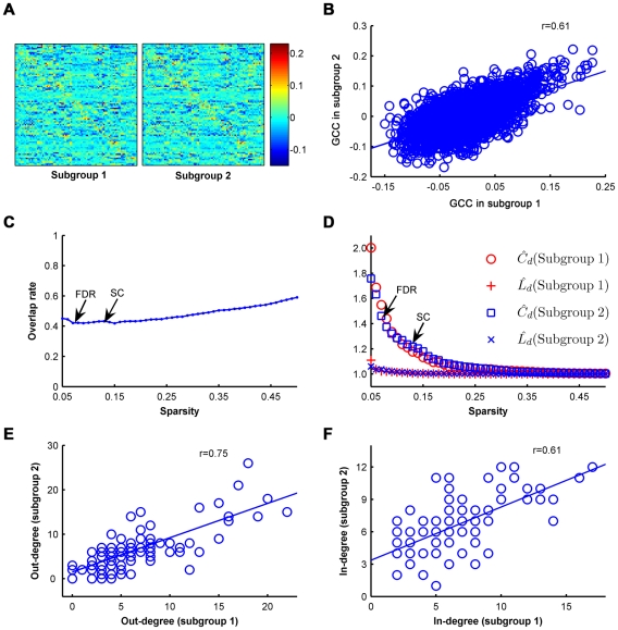
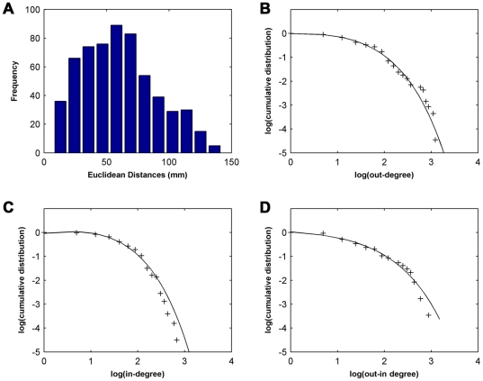
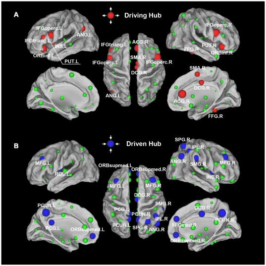
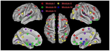
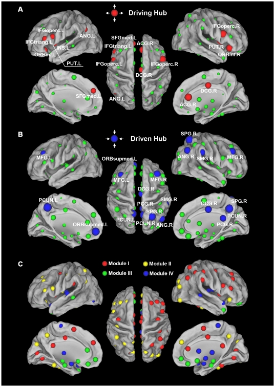
Recently, increasing attention has been focused on the investigation of the human brain connectome that describes the patterns of structural and functional connectivity networks of the human brain. Many studies of the human connectome have demonstrated that the brain network follows a small-world topology with an intrinsically cohesive modular structure and includes several network hubs in the medial parietal regions. However, most of these studies have only focused on undirected connections between regions in which the directions of information flow are not taken into account. How the brain regions causally influence each other and how the directed network of human brain is topologically organized remain largely unknown. Here, we applied linear multivariate Granger causality analysis (GCA) and graph theoretical approaches to a resting-state functional MRI dataset with a large cohort of young healthy participants (n = 86) to explore connectivity patterns of the population-based whole-brain functional directed network. This directed brain network exhibited prominent small-world properties, which obviously improved previous results of functional MRI studies showing weak small-world properties in the directed brain networks in terms of a kernel-based GCA and individual analysis. This brain network also showed significant modular structures associated with 5 well known subsystems: fronto-parietal, visual, paralimbic/limbic, subcortical and primary systems. Importantly, we identified several driving hubs predominantly located in the components of the attentional network (e.g., the inferior frontal gyrus, supplementary motor area, insula and fusiform gyrus) and several driven hubs predominantly located in the components of the default mode network (e.g., the precuneus, posterior cingulate gyrus, medial prefrontal cortex and inferior parietal lobule). Further split-half analyses indicated that our results were highly reproducible between two independent subgroups. The current study demonstrated the directions of spontaneous information flow and causal influences in the directed brain networks, thus providing new insights into our understanding of human brain functional connectome.
最近,人们越来越关注对人类大脑连接组的研究,该研究描述了人类大脑结构和功能连接网络的模式。许多人类连接组的研究表明,大脑网络遵循小世界拓扑结构,具有内在的凝聚模块结构,并包括内侧顶叶区域的几个网络枢纽。然而,大多数这些研究仅关注区域之间的无向连接,而没有考虑信息流的方向。大脑区域如何相互因果影响,以及人类大脑的有向网络在拓扑上是如何组织的,在很大程度上仍然未知。在这里,我们应用线性多元格兰杰因果分析(GCA)和图论方法,对一个具有大量年轻健康参与者(n=86)的静息状态功能磁共振成像数据集进行了分析,以探索基于人群的全脑功能有向网络的连接模式。这个有向大脑网络表现出明显的小世界特性,这明显改进了以前的基于核的 GCA 和个体分析显示有向大脑网络中弱小世界特性的功能磁共振成像研究的结果。该大脑网络还显示出与 5 个已知子系统相关的显著模块结构:额顶叶、视觉、边缘/边缘系统、皮质下和初级系统。重要的是,我们确定了几个主要位于注意力网络组成部分中的驱动枢纽(例如,额下回、辅助运动区、岛叶和梭状回),以及几个主要位于默认模式网络组成部分中的受驱动枢纽(例如,楔前叶、后扣带回、内侧前额叶皮质和下顶叶)。进一步的分组分析表明,我们的结果在两个独立的子组之间具有高度的可重复性。本研究表明了有向大脑网络中自发信息流和因果影响的方向,从而为我们理解人类大脑功能连接组提供了新的视角。