Department of Pathology, University of Sydney, Camperdown, Australia.
J Cell Mol Med. 2012 Aug;16(8):1731-8. doi: 10.1111/j.1582-4934.2011.01434.x.
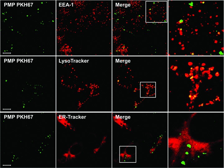
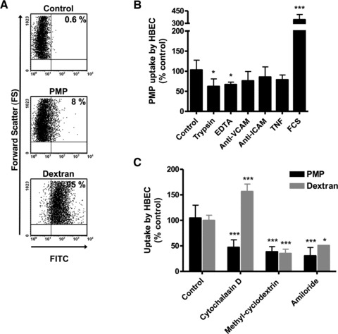
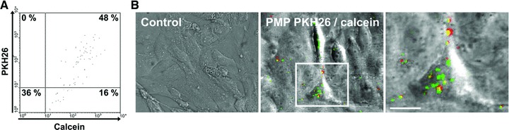
Platelet-derived microparticles (PMP) bind and modify the phenotype of many cell types including endothelial cells. Recently, we showed that PMP were internalized by human brain endothelial cells (HBEC). Here we intend to better characterize the internalization mechanisms of PMP and their intracellular fate. Confocal microscopy analysis of PKH67-labelled PMP distribution in HBEC showed PMP in early endosome antigen 1 positive endosomes and in LysoTracker-labelled lysosomes, confirming a role for endocytosis in PMP internalization. No fusion of calcein-loaded PMP with HBEC membranes was observed. Quantification of PMP endocytosis using flow cytometry revealed that it was partially inhibited by trypsin digestion of PMP surface proteins and by extracellular Ca(2+) chelation by EDTA, suggesting a partial role for receptor-mediated endocytosis in PMP uptake. This endocytosis was independent of endothelial receptors such as intercellular adhesion molecule-1 and vascular cell adhesion molecule-1 and was not increased by tumour necrosis factor stimulation of HBEC. Platelet-derived microparticle internalization was dramatically increased in the presence of decomplemented serum, suggesting a role for PMP opsonin-dependent phagocytosis. Platelet-derived microparticle uptake was greatly diminished by treatment of HBEC with cytochalasin D, an inhibitor of microfilament formation required for both phagocytosis and macropinocytosis, with methyl-β-cyclodextrin that depletes membrane cholesterol needed for macropinocytosis and with amiloride that inhibits the Na(+)/H(+) exchanger involved in macropinocytosis. In conclusion, PMP are taken up by active endocytosis in HBEC, involving mechanisms consistent with both phagocytosis and macropinocytosis. These findings identify new processes by which PMP could modify endothelial cell phenotype and functions.
血小板衍生的微粒 (PMP) 结合并改变许多细胞类型的表型,包括内皮细胞。最近,我们发现 PMP 被人脑血管内皮细胞 (HBEC) 内化。在这里,我们旨在更好地描述 PMP 的内化机制及其细胞内命运。PKH67 标记的 PMP 在 HBEC 中的分布的共聚焦显微镜分析显示 PMP 位于早期内体抗原 1 阳性内体和溶酶体标记的溶酶体中,证实内吞作用在 PMP 内化中起作用。未观察到 calcein 负载的 PMP 与 HBEC 膜融合。使用流式细胞术定量 PMP 内吞作用表明,PMP 表面蛋白的胰蛋白酶消化和 EDTA 螯合细胞外 Ca(2+) 部分抑制了内吞作用,表明 PMP 摄取中存在部分受体介导的内吞作用。这种内吞作用独立于内皮细胞受体,如细胞间黏附分子-1 和血管细胞黏附分子-1,并且不受 HBEC 肿瘤坏死因子刺激的增加。补体失活血清的存在显著增加了血小板衍生的微粒的内化,表明 PMP 调理素依赖性吞噬作用的作用。HBEC 用细胞松弛素 D 处理、微丝形成抑制剂,微丝形成对于吞噬作用和巨胞饮作用都是必需的,用甲基-β-环糊精耗尽巨胞饮作用所需的膜胆固醇,用阿米洛利抑制巨胞饮作用中涉及的 Na(+)/H(+) 交换器,都会大大减少血小板衍生的微粒的摄取。总之,PMP 通过 HBEC 中的主动内吞作用被摄取,涉及与吞噬作用和巨胞饮作用一致的机制。这些发现确定了 PMP 可以改变内皮细胞表型和功能的新过程。