• 文献检索
  • 文档翻译
  • 深度研究
  • 学术资讯
  • Suppr Zotero 插件Zotero 插件
  • 邀请有礼
  • 套餐&价格
  • 历史记录
应用&插件
Suppr Zotero 插件Zotero 插件浏览器插件Mac 客户端Windows 客户端微信小程序
定价
高级版会员购买积分包购买API积分包
服务
文献检索文档翻译深度研究API 文档MCP 服务
关于我们
关于 Suppr公司介绍联系我们用户协议隐私条款
关注我们

Suppr 超能文献

核心技术专利:CN118964589B侵权必究
粤ICP备2023148730 号-1Suppr @ 2026

文献检索

告别复杂PubMed语法,用中文像聊天一样搜索,搜遍4000万医学文献。AI智能推荐,让科研检索更轻松。

立即免费搜索

文件翻译

保留排版,准确专业,支持PDF/Word/PPT等文件格式,支持 12+语言互译。

免费翻译文档

深度研究

AI帮你快速写综述,25分钟生成高质量综述,智能提取关键信息,辅助科研写作。

立即免费体验

基于X射线相衬成像的微小软组织可视化

Micro soft tissues visualization based on x-ray phase-contrast imaging.

作者信息

Zhang Lu, Luo Shuqian

机构信息

School of Biomedical Engineering, Capital Medical University, Beijing, China.

出版信息

Open Med Inform J. 2011;5(Suppl 1):19-25. doi: 10.2174/1874431101105010019. Epub 2011 Jul 27.

DOI:10.2174/1874431101105010019
PMID:21892370
原文链接:https://pmc.ncbi.nlm.nih.gov/articles/PMC3151592/
Abstract

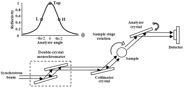
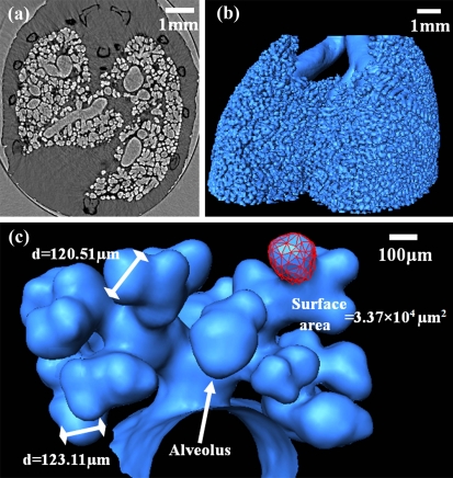
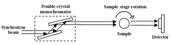
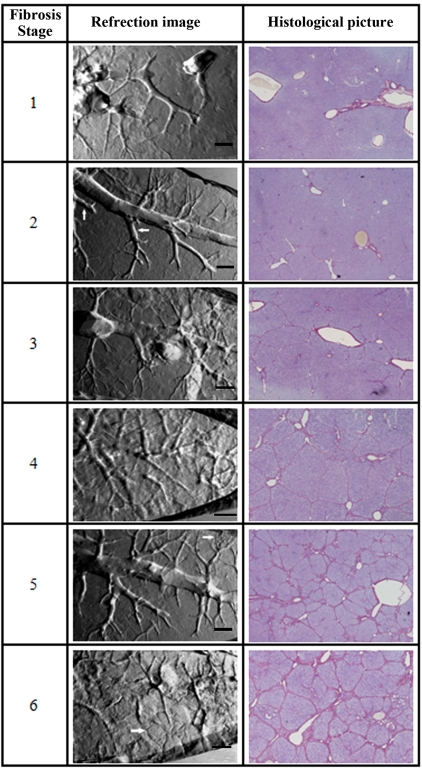
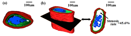
The current imaging methods have a limited ability to visualize microstructures of biological soft tissues. Small lesions cannot be detected at the early stage of the disease. Phase contrast imaging (PCI) is a novel non-invasive imaging technique that can provide high contrast images of soft tissues by the use of X-ray phase shift. It is a new choice in terms of non-invasively revealing soft tissue details. In this study, the lung and hepatic fibrosis models of mice and rats were used to investigate the ability of PCI in microstructures observation of soft tissues. Our results demonstrated that different liver fibrosis stages could be distinguished non-invasively by PCI. The three-dimensional morphology of a segment of blood vessel was constructed. Noteworthy, the blood clot inside the vessel was visualized in three dimensions which provided a precise description of vessel stenosis. Furthermore, the whole lung airways including the alveoli were obtained. We had specifically highlighted its use in the visualization and assessment of the alveoli. To our knowledge, this was the first time for non-invasive alveoli imaging using PCI. This finding may offer a new perspective on the diagnosis of respiratory disease. All the results confirmed that PCI will be a valuable tool in biological soft tissues imaging.

摘要

当前的成像方法在可视化生物软组织微观结构方面能力有限。在疾病早期无法检测到小病变。相衬成像(PCI)是一种新型非侵入性成像技术,它可以通过利用X射线相移提供软组织的高对比度图像。就非侵入性揭示软组织细节而言,它是一种新的选择。在本研究中,使用小鼠和大鼠的肺和肝纤维化模型来研究PCI在软组织微观结构观察方面的能力。我们的结果表明,PCI可以非侵入性地区分不同的肝纤维化阶段。构建了一段血管的三维形态。值得注意的是,血管内的血凝块在三维上可视化,这提供了对血管狭窄的精确描述。此外,获得了包括肺泡在内的整个肺气道。我们特别强调了其在肺泡可视化和评估中的应用。据我们所知,这是首次使用PCI进行非侵入性肺泡成像。这一发现可能为呼吸系统疾病的诊断提供新的视角。所有结果证实,PCI将成为生物软组织成像中的一种有价值的工具。

https://cdn.ncbi.nlm.nih.gov/pmc/blobs/d593/3151592/15405ebc5b44/TOMINFOJ-5-19_F5.jpg
https://cdn.ncbi.nlm.nih.gov/pmc/blobs/d593/3151592/e1f74a5f5fbe/TOMINFOJ-5-19_F1.jpg
https://cdn.ncbi.nlm.nih.gov/pmc/blobs/d593/3151592/c1efd9e6c6f2/TOMINFOJ-5-19_F2.jpg
https://cdn.ncbi.nlm.nih.gov/pmc/blobs/d593/3151592/084671d90c09/TOMINFOJ-5-19_F3.jpg
https://cdn.ncbi.nlm.nih.gov/pmc/blobs/d593/3151592/4d76624bb5a7/TOMINFOJ-5-19_F4.jpg
https://cdn.ncbi.nlm.nih.gov/pmc/blobs/d593/3151592/15405ebc5b44/TOMINFOJ-5-19_F5.jpg

相似文献

1
Micro soft tissues visualization based on x-ray phase-contrast imaging.基于X射线相衬成像的微小软组织可视化
Open Med Inform J. 2011;5(Suppl 1):19-25. doi: 10.2174/1874431101105010019. Epub 2011 Jul 27.
2
Non-invasive microstructure and morphology investigation of the mouse lung: qualitative description and quantitative measurement.无创性微观结构和形态学研究的老鼠肺:定性描述和定量测量。
PLoS One. 2011 Feb 25;6(2):e17400. doi: 10.1371/journal.pone.0017400.
3
Noninvasive visualization of microvessels using diffraction enhanced imaging.利用衍射增强成像技术进行微血管无创可视化。
Eur J Radiol. 2011 Oct;80(1):158-62. doi: 10.1016/j.ejrad.2010.08.019. Epub 2010 Sep 15.
4
Improved middle-ear soft-tissue visualization using synchrotron radiation phase-contrast imaging.使用同步辐射相衬成像改善中耳软组织可视化。
Hear Res. 2017 Oct;354:1-8. doi: 10.1016/j.heares.2017.08.001. Epub 2017 Aug 5.
5
Three-dimensional visualization of the microvasculature of bile duct ligation-induced liver fibrosis in rats by x-ray phase-contrast imaging computed tomography.通过X射线相衬成像计算机断层扫描对胆管结扎诱导的大鼠肝纤维化微血管进行三维可视化
Sci Rep. 2015 Jul 27;5:11500. doi: 10.1038/srep11500.
6
Microcomputed tomography with diffraction-enhanced imaging for morphologic characterization and quantitative evaluation of microvessel of hepatic fibrosis in rats.微计算机断层扫描结合衍射增强成像用于大鼠肝纤维化微血管的形态学表征和定量评估
PLoS One. 2013 Oct 21;8(10):e78176. doi: 10.1371/journal.pone.0078176. eCollection 2013.
7
X-ray diffraction enhanced imaging as a novel method to visualize low-density scaffolds in soft tissue engineering.X 射线衍射增强成像作为一种新方法,可用于可视化软组织工程中的低密度支架。
Tissue Eng Part C Methods. 2011 Nov;17(11):1071-80. doi: 10.1089/ten.tec.2011.0102. Epub 2011 Aug 26.
8
A micro-tomography method based on X-ray diffraction enhanced imaging for the visualization of micro-organs and soft tissues.一种基于X射线衍射增强成像的显微断层扫描方法,用于微器官和软组织的可视化。
Comput Med Imaging Graph. 2006 Sep-Oct;30(6-7):339-47. doi: 10.1016/j.compmedimag.2006.09.003. Epub 2006 Oct 18.
9
High-resolution visualization of airspace structures in intact mice via synchrotron phase-contrast X-ray imaging (PCXI).利用同步辐射相衬 X 射线成像(PCXI)技术对完整小鼠的气腔结构进行高分辨率可视化。
J Anat. 2008 Aug;213(2):217-27. doi: 10.1111/j.1469-7580.2008.00950.x.
10
Potential of propagation-based synchrotron X-ray phase-contrast computed tomography for cardiac tissue engineering.基于传播的同步加速器X射线相衬计算机断层扫描在心脏组织工程中的潜力。
J Synchrotron Radiat. 2017 Jul 1;24(Pt 4):842-853. doi: 10.1107/S1600577517006208. Epub 2017 May 18.

引用本文的文献

1
Cartilage Tissue Engineering Approaches Need to Assess Fibrocartilage When Hydrogel Constructs Are Mechanically Loaded.当水凝胶构建体受到机械加载时,软骨组织工程方法需要评估纤维软骨。
Front Bioeng Biotechnol. 2022 Jan 12;9:787538. doi: 10.3389/fbioe.2021.787538. eCollection 2021.
2
Computed Tomography as a Characterization Tool for Engineered Scaffolds with Biomedical Applications.计算机断层扫描作为具有生物医学应用的工程支架的表征工具。
Materials (Basel). 2021 Nov 10;14(22):6763. doi: 10.3390/ma14226763.
3
Microdissection of Human Esophagogastric Junction Wall with Phase-contrast X-ray CT Imaging.

本文引用的文献

1
Non-invasive microstructure and morphology investigation of the mouse lung: qualitative description and quantitative measurement.无创性微观结构和形态学研究的老鼠肺:定性描述和定量测量。
PLoS One. 2011 Feb 25;6(2):e17400. doi: 10.1371/journal.pone.0017400.
2
Noninvasive visualization of microvessels using diffraction enhanced imaging.利用衍射增强成像技术进行微血管无创可视化。
Eur J Radiol. 2011 Oct;80(1):158-62. doi: 10.1016/j.ejrad.2010.08.019. Epub 2010 Sep 15.
3
Comparison of two quantitative methods of discerning airspace enlargement in smoke-exposed mice.
利用相衬X射线计算机断层成像对人食管胃交界壁进行显微解剖
Sci Rep. 2015 Sep 8;5:13831. doi: 10.1038/srep13831.
4
Phase-contrast X-ray CT imaging of esophagus and esophageal carcinoma.食管及食管癌的相衬X线计算机断层成像
Sci Rep. 2014 Jun 18;4:5332. doi: 10.1038/srep05332.
5
Imaging challenges in biomaterials and tissue engineering.生物材料和组织工程中的成像挑战。
Biomaterials. 2013 Sep;34(28):6615-30. doi: 10.1016/j.biomaterials.2013.05.033. Epub 2013 Jun 13.
比较两种定量方法识别烟熏暴露小鼠的气腔扩大。
PLoS One. 2009 Aug 18;4(8):e6670. doi: 10.1371/journal.pone.0006670.
4
Endophenotypes in normal brain morphology and Alzheimer's disease: a review.正常脑形态学和阿尔茨海默病的内表型:综述。
Neuroscience. 2009 Nov 24;164(1):174-90. doi: 10.1016/j.neuroscience.2009.04.006. Epub 2009 Apr 9.
5
High-resolution visualization of airspace structures in intact mice via synchrotron phase-contrast X-ray imaging (PCXI).利用同步辐射相衬 X 射线成像(PCXI)技术对完整小鼠的气腔结构进行高分辨率可视化。
J Anat. 2008 Aug;213(2):217-27. doi: 10.1111/j.1469-7580.2008.00950.x.
6
Evaluation of microvascular endothelial function in patients with type 1 diabetes using laser-Doppler perfusion monitoring: which method to choose?使用激光多普勒灌注监测评估1型糖尿病患者的微血管内皮功能:应选择哪种方法?
Microvasc Res. 2008 Aug;76(2):132-3. doi: 10.1016/j.mvr.2008.04.003. Epub 2008 Apr 25.
7
Non-invasive evaluation of liver fibrosis using transient elastography.使用瞬时弹性成像技术对肝纤维化进行无创评估。
J Hepatol. 2008 May;48(5):835-47. doi: 10.1016/j.jhep.2008.02.008. Epub 2008 Feb 26.
8
Microvascular tumor invasion, tumor size and Fuhrman grade: a pathological triad for prognostic evaluation of renal cell carcinoma.微血管肿瘤浸润、肿瘤大小与富尔曼分级:肾细胞癌预后评估的病理三联征
J Urol. 2007 Aug;178(2):425-8; discussion 428. doi: 10.1016/j.juro.2007.03.128. Epub 2007 Jun 11.
9
Adverse reactions to contrast media: an analysis from spontaneous reporting data.造影剂不良反应:基于自发报告数据的分析
Pharmacol Res. 2007 Jul;56(1):35-41. doi: 10.1016/j.phrs.2007.03.003. Epub 2007 Mar 25.
10
Simvastatin prevents coronary microvascular remodeling in renovascular hypertensive pigs.辛伐他汀可预防肾血管性高血压猪的冠状动脉微血管重塑。
J Am Soc Nephrol. 2007 Apr;18(4):1209-17. doi: 10.1681/ASN.2006090976. Epub 2007 Mar 7.