• 文献检索
  • 文档翻译
  • 深度研究
  • 学术资讯
  • Suppr Zotero 插件Zotero 插件
  • 邀请有礼
  • 套餐&价格
  • 历史记录
应用&插件
Suppr Zotero 插件Zotero 插件浏览器插件Mac 客户端Windows 客户端微信小程序
定价
高级版会员购买积分包购买API积分包
服务
文献检索文档翻译深度研究API 文档MCP 服务
关于我们
关于 Suppr公司介绍联系我们用户协议隐私条款
关注我们

Suppr 超能文献

核心技术专利:CN118964589B侵权必究
粤ICP备2023148730 号-1Suppr @ 2026

文献检索

告别复杂PubMed语法,用中文像聊天一样搜索,搜遍4000万医学文献。AI智能推荐,让科研检索更轻松。

立即免费搜索

文件翻译

保留排版,准确专业,支持PDF/Word/PPT等文件格式,支持 12+语言互译。

免费翻译文档

深度研究

AI帮你快速写综述,25分钟生成高质量综述,智能提取关键信息,辅助科研写作。

立即免费体验

食管及食管癌的相衬X线计算机断层成像

Phase-contrast X-ray CT imaging of esophagus and esophageal carcinoma.

作者信息

Zhang Jianfa, Tian Dongping, Lin Runhua, Zhou Guangzhao, Peng Guanyun, Su Min

机构信息

First Affiliated Hospital of Shantou University Medical College, Shantou, Guangdong, People's Republic of China.

1] Institute of Clinical Pathology & Department of Pathology, Shantou University Medical College, Shantou, Guangdong, People's Republic of China [2] The Judicial Critical Center, Shantou University Medical College, Shantou, Guangdong, People's Republic of China.

出版信息

Sci Rep. 2014 Jun 18;4:5332. doi: 10.1038/srep05332.

DOI:10.1038/srep05332
PMID:24939041
原文链接:https://pmc.ncbi.nlm.nih.gov/articles/PMC4061548/
Abstract

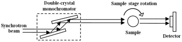
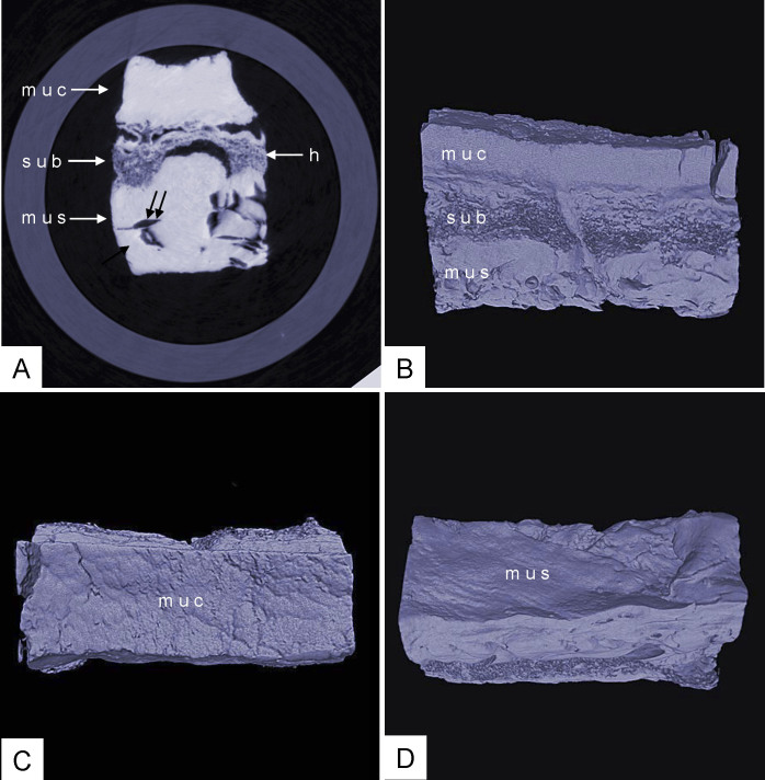
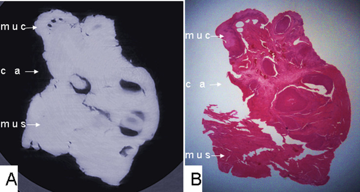
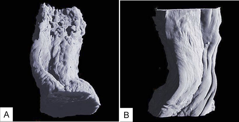
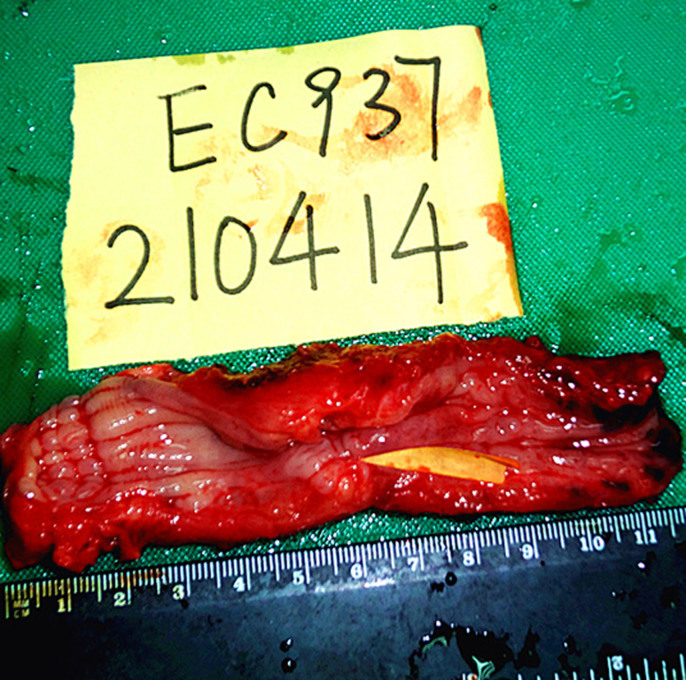
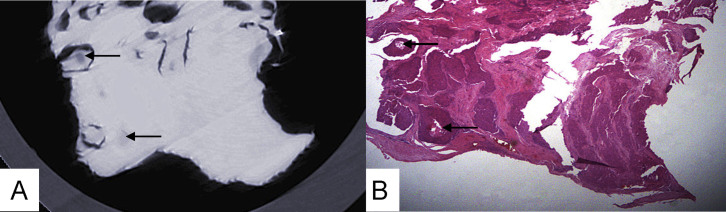
The electron density resolution is 1000 times higher for synchrotron-radiation phase-contrast CT imaging than conventional X-ray absorption imaging in light elements, with which high-resolution X-ray imaging of biological soft tissue can be achieved. In the present study, we used phase-contrast X-ray CT to investigate human resected esophagus and esophageal carcinoma specimens. This technology revealed the three-layer structure of the esophageal wall-- mucous, submucosa and muscular layers. The mucous and muscular layers were clearly separated by a loose submucosa layer with a honeycomb appearance. The surface of the mucous layer was smooth. In esophageal carcinoma, because of tumor tissue infiltration, the submucosa layer was absent, which indicated destruction of the submucosa. The boundary between normal tissue and tumor was comparatively fuzzy, the three-layer structure of the esophageal wall was indistinct. The surface of the mucous layer was rugose. The technology might be helpful in tumor staging of esophageal carcinoma.

摘要

在轻元素中,同步辐射相衬CT成像的电子密度分辨率比传统X射线吸收成像高1000倍,利用这一点可以实现生物软组织的高分辨率X射线成像。在本研究中,我们使用相衬X射线CT对人切除的食管和食管癌标本进行研究。这项技术揭示了食管壁的三层结构——黏膜层、黏膜下层和肌层。黏膜层和肌层被具有蜂窝状外观的疏松黏膜下层清晰分隔。黏膜层表面光滑。在食管癌中,由于肿瘤组织浸润,黏膜下层缺失,这表明黏膜下层遭到破坏。正常组织与肿瘤之间的边界相对模糊,食管壁的三层结构不清晰。黏膜层表面粗糙。这项技术可能有助于食管癌的肿瘤分期。

https://cdn.ncbi.nlm.nih.gov/pmc/blobs/bc3b/4061548/5b0b8fa06e35/srep05332-f8.jpg
https://cdn.ncbi.nlm.nih.gov/pmc/blobs/bc3b/4061548/7202097e7971/srep05332-f1.jpg
https://cdn.ncbi.nlm.nih.gov/pmc/blobs/bc3b/4061548/c8f4ac6133ea/srep05332-f2.jpg
https://cdn.ncbi.nlm.nih.gov/pmc/blobs/bc3b/4061548/8f702c21f3d0/srep05332-f3.jpg
https://cdn.ncbi.nlm.nih.gov/pmc/blobs/bc3b/4061548/84ec393e2494/srep05332-f4.jpg
https://cdn.ncbi.nlm.nih.gov/pmc/blobs/bc3b/4061548/13c272fdca51/srep05332-f5.jpg
https://cdn.ncbi.nlm.nih.gov/pmc/blobs/bc3b/4061548/a95125524c04/srep05332-f6.jpg
https://cdn.ncbi.nlm.nih.gov/pmc/blobs/bc3b/4061548/ffa45c574c4e/srep05332-f7.jpg
https://cdn.ncbi.nlm.nih.gov/pmc/blobs/bc3b/4061548/5b0b8fa06e35/srep05332-f8.jpg

相似文献

1
Phase-contrast X-ray CT imaging of esophagus and esophageal carcinoma.食管及食管癌的相衬X线计算机断层成像
Sci Rep. 2014 Jun 18;4:5332. doi: 10.1038/srep05332.
2
Microdissection of Human Esophagogastric Junction Wall with Phase-contrast X-ray CT Imaging.利用相衬X射线计算机断层成像对人食管胃交界壁进行显微解剖
Sci Rep. 2015 Sep 8;5:13831. doi: 10.1038/srep13831.
3
Synchrotron-radiation phase-contrast imaging of human stomach and gastric cancer: in vitro studies.同步辐射相衬成像在人胃和胃癌中的应用:体外研究。
J Synchrotron Radiat. 2012 May;19(Pt 3):319-22. doi: 10.1107/S090904951200310X. Epub 2012 Mar 15.
4
High contrast microstructural visualization of natural acellular matrices by means of phase-based x-ray tomography.通过基于相位的X射线断层扫描对天然无细胞基质进行高对比度微观结构可视化。
Sci Rep. 2015 Dec 14;5:18156. doi: 10.1038/srep18156.
5
Esophageal imaging by MR and CT: study of normal anatomy and neoplasms.食管的磁共振成像和计算机断层扫描:正常解剖结构与肿瘤的研究
Radiology. 1985 Sep;156(3):727-31. doi: 10.1148/radiology.156.3.4023234.
6
[The value of computed tomography for the pretherapeutic staging of esophageal cancer].[计算机断层扫描在食管癌治疗前分期中的价值]
Radiol Diagn (Berl). 1989;30(2):111-7.
7
Visualization Ability of Phase-Contrast Synchrotron-Based X-Ray Imaging Using an X-Ray Interferometer in Soft Tissue Tumors.基于相衬同步辐射 X 射线干涉仪的软组织肿瘤 X 射线成像的可视化能力。
Technol Cancer Res Treat. 2021 Jan-Dec;20:15330338211010121. doi: 10.1177/15330338211010121.
8
[Esophagus carcinoma--progress in staging with thin layer spiral CT?].[食管癌——薄层螺旋CT在分期方面的进展?]
Rontgenpraxis. 1998;51(4):130-4.
9
Translation of atherosclerotic plaque phase-contrast CT imaging from synchrotron radiation to a conventional lab-based X-ray source.从同步辐射到常规实验室基于 X 射线源的动脉粥样硬化斑块相衬 CT 成像的转换。
PLoS One. 2013 Sep 9;8(9):e73513. doi: 10.1371/journal.pone.0073513. eCollection 2013.
10
Dedicated multi-detector CT of the esophagus: spectrum of diseases.食管专用多排CT:疾病谱
Abdom Imaging. 2009 Jan-Feb;34(1):3-18. doi: 10.1007/s00261-007-9290-5.

引用本文的文献

1
Unveiling Another Dimension: Advanced Visualization of Cancer Invasion and Metastasis via Micro-CT Imaging.揭示另一个维度:通过微型计算机断层扫描成像对癌症侵袭和转移进行高级可视化
Cancers (Basel). 2025 Mar 28;17(7):1139. doi: 10.3390/cancers17071139.
2
T staging esophageal tumors with x rays.用X线对食管癌进行T分期
Optica. 2024 Apr 19;11(4):569-576. doi: 10.1364/OPTICA.501948. eCollection 2024 Apr 20.
3
High-Resolution Phase-Contrast Tomography on Human Collagenous Tissues: A Comprehensive Review.高分辨率相衬层析术在人体胶原组织中的应用:全面综述。

本文引用的文献

1
Use of holmium:YAG laser in posterior urethral valves: another method of fulguration.钬激光在治疗后尿道瓣膜中的应用:另一种烧灼方法。
J Pediatr Urol. 2013 Dec;9(6 Pt B):1093-7. doi: 10.1016/j.jpurol.2013.03.015. Epub 2013 May 6.
2
Molecular pathological diagnosis for early esophageal cancer in Kazakh patients.哈萨克族食管癌患者早期分子病理诊断
Oncol Lett. 2012 Mar;3(3):549-553. doi: 10.3892/ol.2011.547. Epub 2011 Dec 30.
3
Synchrotron-radiation phase-contrast imaging of human stomach and gastric cancer: in vitro studies.
Tomography. 2023 Nov 27;9(6):2116-2133. doi: 10.3390/tomography9060166.
4
Visualising the 3D microstructure of stained and native intervertebral discs using X-ray microtomography.利用 X 射线显微断层扫描技术对染色和天然椎间盘中的 3D 微观结构进行可视化。
Sci Rep. 2017 Nov 24;7(1):16279. doi: 10.1038/s41598-017-16354-w.
5
3D visualization of the lumbar facet joint after degeneration using propagation phase contrast micro-tomography.使用传播相位对比显微断层扫描技术对退变后腰椎小关节进行三维可视化。
Sci Rep. 2016 Feb 24;6:21838. doi: 10.1038/srep21838.
6
Phase-Contrast Micro-Computed Tomography Measurements of the Intraocular Pressure-Induced Deformation of the Porcine Lamina Cribrosa.眼压诱导的猪筛板变形的相衬显微计算机断层扫描测量
IEEE Trans Med Imaging. 2016 Apr;35(4):988-99. doi: 10.1109/TMI.2015.2504440. Epub 2015 Nov 30.
7
X-ray phase-contrast tomography for high-spatial-resolution zebrafish muscle imaging.用于高空间分辨率斑马鱼肌肉成像的X射线相衬断层扫描术
Sci Rep. 2015 Nov 13;5:16625. doi: 10.1038/srep16625.
8
Microdissection of Human Esophagogastric Junction Wall with Phase-contrast X-ray CT Imaging.利用相衬X射线计算机断层成像对人食管胃交界壁进行显微解剖
Sci Rep. 2015 Sep 8;5:13831. doi: 10.1038/srep13831.
9
Three-dimensional visualization of the microvasculature of bile duct ligation-induced liver fibrosis in rats by x-ray phase-contrast imaging computed tomography.通过X射线相衬成像计算机断层扫描对胆管结扎诱导的大鼠肝纤维化微血管进行三维可视化
Sci Rep. 2015 Jul 27;5:11500. doi: 10.1038/srep11500.
同步辐射相衬成像在人胃和胃癌中的应用:体外研究。
J Synchrotron Radiat. 2012 May;19(Pt 3):319-22. doi: 10.1107/S090904951200310X. Epub 2012 Mar 15.
4
Micro soft tissues visualization based on x-ray phase-contrast imaging.基于X射线相衬成像的微小软组织可视化
Open Med Inform J. 2011;5(Suppl 1):19-25. doi: 10.2174/1874431101105010019. Epub 2011 Jul 27.
5
A novel tumor suppressor gene ECRG4 interacts directly with TMPRSS11A (ECRG1) to inhibit cancer cell growth in esophageal carcinoma.一个新的抑癌基因 ECRG4 与 TMPRSS11A(ECRG1)直接相互作用,抑制食管癌中的癌细胞生长。
BMC Cancer. 2011 Feb 3;11:52. doi: 10.1186/1471-2407-11-52.
6
Multimodality therapy for esophageal cancer.食管癌的多模态治疗。
Oncology (Williston Park). 2010 Nov 15;24(12):1134-8.
7
Epidemiology and pathogenesis of esophageal cancer.食管癌的流行病学与发病机制
Semin Radiat Oncol. 2007 Jan;17(1):2-9. doi: 10.1016/j.semradonc.2006.09.003.
8
Phase-contrast X-ray imaging with a large monolithic X-ray interferometer.使用大型整体式X射线干涉仪的相衬X射线成像。
J Synchrotron Radiat. 2000 Jul 1;7(Pt 4):280-2. doi: 10.1107/S0909049500004295.
9
Global cancer statistics, 2002.2002年全球癌症统计数据。
CA Cancer J Clin. 2005 Mar-Apr;55(2):74-108. doi: 10.3322/canjclin.55.2.74.
10
Accuracy of staging in early oesophageal cancer using high resolution endoscopy and high resolution endosonography: a comparative, prospective, and blinded trial.使用高分辨率内镜检查和高分辨率超声内镜对早期食管癌进行分期的准确性:一项对比性、前瞻性、双盲试验。
Gut. 2004 May;53(5):634-40. doi: 10.1136/gut.2003.029421.