Department of Radiation Therapy, Charité Medical Center, Berlin, Germany.
Radiat Oncol. 2011 Sep 5;6:107. doi: 10.1186/1748-717X-6-107.
To assess brachytherapy catheter positioning accuracy and to evaluate the effects of prolonged irradiation time on the tolerance dose of normal liver parenchyma following single-fraction irradiation with 192Ir.
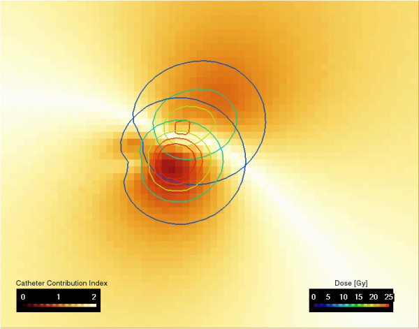
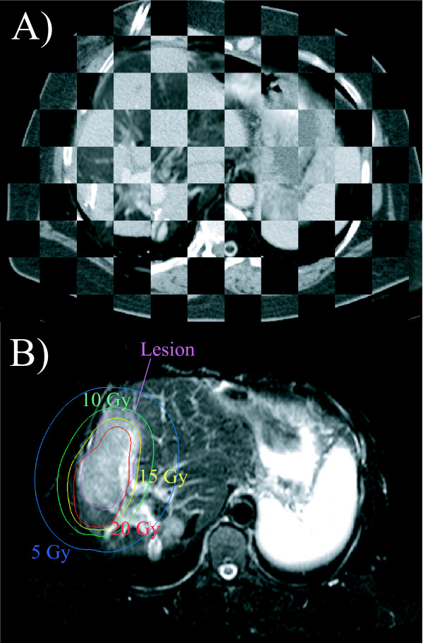
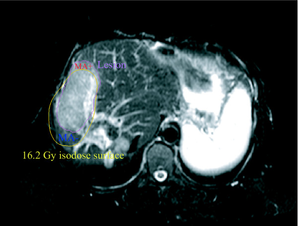
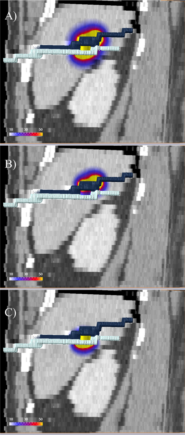
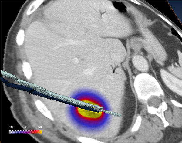
Fifty patients with 76 malignant liver tumors treated by computed tomography (CT)-guided high-dose-rate brachytherapy (HDR-BT) were included in the study. The prescribed radiation dose was delivered by 1 - 11 catheters with exposure times in the range of 844 - 4432 seconds. Magnetic resonance imaging (MRI) datasets for assessing irradiation effects on normal liver tissue, edema, and hepatocyte dysfunction, obtained 6 and 12 weeks after HDR-BT, were merged with 3D dosimetry data. The isodose of the treatment plan covering the same volume as the irradiation effect was taken as a surrogate for the liver tissue tolerance dose. Catheter positioning accuracy was assessed by calculating the shift between the 3D center coordinates of the irradiation effect volume and the tolerance dose volume for 38 irradiation effects in 30 patients induced by catheters implanted in nearly parallel arrangement. Effects of prolonged irradiation were assessed in areas where the irradiation effect volume and tolerance dose volume did not overlap (mismatch areas) by using a catheter contribution index. This index was calculated for 48 irradiation effects induced by at least two catheters in 44 patients.
Positioning accuracy of the brachytherapy catheters was 5-6 mm. The orthogonal and axial shifts between the center coordinates of the irradiation effect volume and the tolerance dose volume in relation to the direction vector of catheter implantation were highly correlated and in first approximation identically in the T1-w and T2-w MRI sequences (p = 0.003 and p < 0.001, respectively), as were the shifts between 6 and 12 weeks examinations (p = 0.001 and p = 0.004, respectively). There was a significant shift of the irradiation effect towards the catheter entry site compared with the planned dose distribution (p < 0.005). Prolonged treatment time increases the normal tissue tolerance dose. Here, the catheter contribution indices indicated a lower tolerance dose of the liver parenchyma in areas with prolonged irradiation (p < 0.005).
Positioning accuracy of brachytherapy catheters is sufficient for clinical practice. Reduced tolerance dose in areas exposed to prolonged irradiation is contradictory to results published in the current literature. Effects of prolonged dose administration on the liver tolerance dose for treatment times of up to 60 minutes per HDR-BT session are not pronounced compared to effects of positioning accuracy of the brachytherapy catheters and are therefore of minor importance in treatment planning.
评估近距离放射治疗导管定位的准确性,并评价单次 192Ir 照射后,延长照射时间对正常肝实质耐受剂量的影响。
本研究纳入了 50 例 76 个恶性肝脏肿瘤患者,采用 CT 引导下高剂量率近距离放射治疗(HDR-BT)。规定的照射剂量由 1-11 根导管提供,照射时间范围为 844-4432 秒。在 HDR-BT 后 6 周和 12 周,获得了用于评估正常肝组织、水肿和肝细胞功能障碍的磁共振成像(MRI)数据集,并与 3D 剂量数据合并。将照射效果覆盖的相同体积的治疗计划等剂量线作为肝组织耐受剂量的替代物。通过计算 30 例患者中近平行排列植入的导管所诱导的 38 个照射效果的 3D 中心坐标的移位,评估导管定位的准确性。通过使用导管贡献指数,在照射效果体积和耐受剂量体积不重叠的区域(失配区域)评估延长照射的效果。该指数是在 44 例患者中至少两根导管诱导的 48 个照射效果中计算得出的。
近距离放射治疗导管的定位精度为 5-6 毫米。照射效果体积和耐受剂量体积的中心坐标与导管植入方向向量之间的正交和轴向移位在 T1-w 和 T2-w MRI 序列中高度相关,且在第一近似值中完全相同(p = 0.003 和 p <0.001),6 周和 12 周检查之间的移位也是如此(p = 0.001 和 p = 0.004)。与计划剂量分布相比,照射效果向导管进入部位发生显著移位(p <0.005)。延长治疗时间会增加正常组织的耐受剂量。在此,导管贡献指数表明,在延长照射的区域,肝实质的耐受剂量较低(p <0.005)。
近距离放射治疗导管的定位精度足以满足临床实践的需要。与当前文献中发表的结果相反,在延长照射区域,耐受剂量降低。与近距离放射治疗导管的定位精度相比,单次 HDR-BT 治疗时间长达 60 分钟内延长剂量给药对肝耐受剂量的影响并不明显,因此在治疗计划中并不重要。