• 文献检索
  • 文档翻译
  • 深度研究
  • 学术资讯
  • Suppr Zotero 插件Zotero 插件
  • 邀请有礼
  • 套餐&价格
  • 历史记录
应用&插件
Suppr Zotero 插件Zotero 插件浏览器插件Mac 客户端Windows 客户端微信小程序
定价
高级版会员购买积分包购买API积分包
服务
文献检索文档翻译深度研究API 文档MCP 服务
关于我们
关于 Suppr公司介绍联系我们用户协议隐私条款
关注我们

Suppr 超能文献

核心技术专利:CN118964589B侵权必究
粤ICP备2023148730 号-1Suppr @ 2026

文献检索

告别复杂PubMed语法,用中文像聊天一样搜索,搜遍4000万医学文献。AI智能推荐,让科研检索更轻松。

立即免费搜索

文件翻译

保留排版,准确专业,支持PDF/Word/PPT等文件格式,支持 12+语言互译。

免费翻译文档

深度研究

AI帮你快速写综述,25分钟生成高质量综述,智能提取关键信息,辅助科研写作。

立即免费体验

用于磁共振图像中脑肿瘤自动分割的图像处理软件的开发。

Development of image-processing software for automatic segmentation of brain tumors in MR images.

作者信息

Vijayakumar C, Gharpure Damayanti Chandrashekhar

机构信息

Department of Radiodiagnosis and Imaging, Armed Forces Medical College, Pune, Maharashtra, India.

出版信息

J Med Phys. 2011 Jul;36(3):147-58. doi: 10.4103/0971-6203.83481.

DOI:10.4103/0971-6203.83481
PMID:21897560
原文链接:https://pmc.ncbi.nlm.nih.gov/articles/PMC3159221/
Abstract

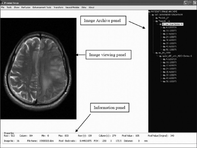
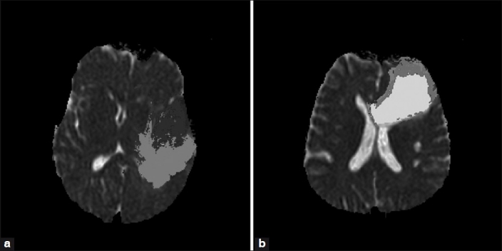
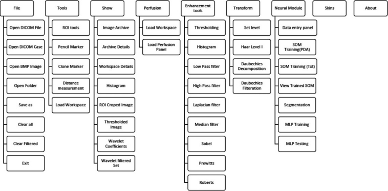
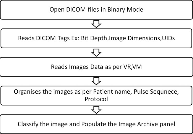
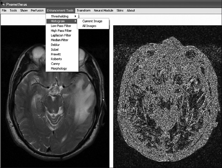
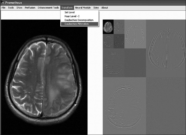
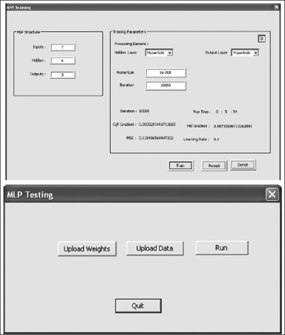
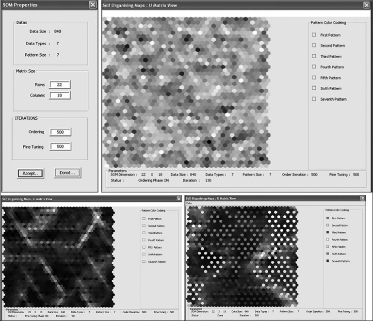
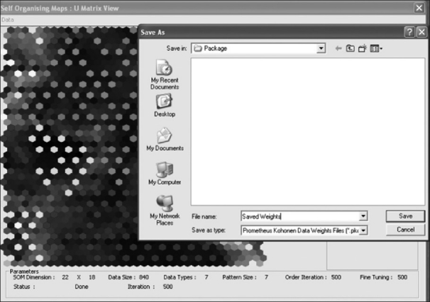
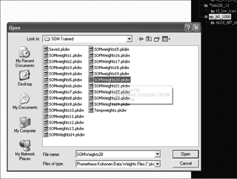
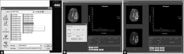
Most of the commercially available software for brain tumor segmentation have limited functionality and frequently lack the careful validation that is required for clinical studies. We have developed an image-analysis software package called 'Prometheus,' which performs neural system-based segmentation operations on MR images using pre-trained information. The software also has the capability to improve its segmentation performance by using the training module of the neural system. The aim of this article is to present the design and modules of this software. The segmentation module of Prometheus can be used primarily for image analysis in MR images. Prometheus was validated against manual segmentation by a radiologist and its mean sensitivity and specificity was found to be 85.71±4.89% and 93.2±2.87%, respectively. Similarly, the mean segmentation accuracy and mean correspondence ratio was found to be 92.35±3.37% and 0.78±0.046, respectively.

摘要

大多数市售的脑肿瘤分割软件功能有限,且常常缺乏临床研究所需的严格验证。我们开发了一个名为“普罗米修斯”的图像分析软件包,它利用预训练信息对磁共振成像(MR图像)执行基于神经系统的分割操作。该软件还能够通过使用神经系统的训练模块来提高其分割性能。本文的目的是介绍该软件的设计和模块。普罗米修斯的分割模块主要可用于MR图像的分析。通过与放射科医生的手动分割进行对比验证,发现普罗米修斯的平均灵敏度和特异性分别为85.71±4.89%和93.2±2.87%。同样,平均分割准确率和平均对应率分别为92.35±3.37%和0.78±0.046。

https://cdn.ncbi.nlm.nih.gov/pmc/blobs/2a39/3159221/b028a78513b0/JMP-36-147-g021.jpg
https://cdn.ncbi.nlm.nih.gov/pmc/blobs/2a39/3159221/37ccb1dceb79/JMP-36-147-g001.jpg
https://cdn.ncbi.nlm.nih.gov/pmc/blobs/2a39/3159221/0a2666315992/JMP-36-147-g002.jpg
https://cdn.ncbi.nlm.nih.gov/pmc/blobs/2a39/3159221/5bb8510e3282/JMP-36-147-g003.jpg
https://cdn.ncbi.nlm.nih.gov/pmc/blobs/2a39/3159221/8c67a05adba8/JMP-36-147-g004.jpg
https://cdn.ncbi.nlm.nih.gov/pmc/blobs/2a39/3159221/9754e65bc5f4/JMP-36-147-g005.jpg
https://cdn.ncbi.nlm.nih.gov/pmc/blobs/2a39/3159221/a93baa32a579/JMP-36-147-g006.jpg
https://cdn.ncbi.nlm.nih.gov/pmc/blobs/2a39/3159221/adc1e904cc58/JMP-36-147-g007.jpg
https://cdn.ncbi.nlm.nih.gov/pmc/blobs/2a39/3159221/3a23bb8fa4a9/JMP-36-147-g009.jpg
https://cdn.ncbi.nlm.nih.gov/pmc/blobs/2a39/3159221/220bc135e4d8/JMP-36-147-g013.jpg
https://cdn.ncbi.nlm.nih.gov/pmc/blobs/2a39/3159221/f00bcb51f9ee/JMP-36-147-g014.jpg
https://cdn.ncbi.nlm.nih.gov/pmc/blobs/2a39/3159221/7e167904f955/JMP-36-147-g015.jpg
https://cdn.ncbi.nlm.nih.gov/pmc/blobs/2a39/3159221/bb60812f47d8/JMP-36-147-g016.jpg
https://cdn.ncbi.nlm.nih.gov/pmc/blobs/2a39/3159221/48527c0e0dc0/JMP-36-147-g017.jpg
https://cdn.ncbi.nlm.nih.gov/pmc/blobs/2a39/3159221/1a53f684cb4c/JMP-36-147-g018.jpg
https://cdn.ncbi.nlm.nih.gov/pmc/blobs/2a39/3159221/9849874ce510/JMP-36-147-g019.jpg
https://cdn.ncbi.nlm.nih.gov/pmc/blobs/2a39/3159221/9e7bd6f822f2/JMP-36-147-g020.jpg
https://cdn.ncbi.nlm.nih.gov/pmc/blobs/2a39/3159221/b028a78513b0/JMP-36-147-g021.jpg

相似文献

1
Development of image-processing software for automatic segmentation of brain tumors in MR images.用于磁共振图像中脑肿瘤自动分割的图像处理软件的开发。
J Med Phys. 2011 Jul;36(3):147-58. doi: 10.4103/0971-6203.83481.
2
Segmentation of organs-at-risks in head and neck CT images using convolutional neural networks.使用卷积神经网络对头颈部CT图像中的危险器官进行分割。
Med Phys. 2017 Feb;44(2):547-557. doi: 10.1002/mp.12045.
3
Design and validation of Segment--freely available software for cardiovascular image analysis.设计和验证——用于心血管图像分析的免费软件段。
BMC Med Imaging. 2010 Jan 11;10:1. doi: 10.1186/1471-2342-10-1.
4
A novel framework for MR image segmentation and quantification by using MedGA.利用 MedGA 实现磁共振图像分割和定量分析的新框架
Comput Methods Programs Biomed. 2019 Jul;176:159-172. doi: 10.1016/j.cmpb.2019.04.016. Epub 2019 Apr 17.
5
Deep HT: A deep neural network for diagnose on MR images of tumors of the hand.深度 HT:用于手部肿瘤磁共振图像诊断的深度神经网络。
PLoS One. 2020 Aug 14;15(8):e0237606. doi: 10.1371/journal.pone.0237606. eCollection 2020.
6
Semi-automatic segmentation software for quantitative clinical brain glioblastoma evaluation.半自动分割软件用于定量临床脑胶质母细胞瘤评估。
Acad Radiol. 2012 Aug;19(8):977-85. doi: 10.1016/j.acra.2012.03.026. Epub 2012 May 15.
7
Automatic segmentation of thoracic and pelvic CT images for radiotherapy planning using implicit anatomic knowledge and organ-specific segmentation strategies.利用隐式解剖学知识和器官特异性分割策略对胸部和盆腔CT图像进行自动分割以用于放射治疗计划。
Phys Med Biol. 2008 Mar 21;53(6):1751-71. doi: 10.1088/0031-9155/53/6/017. Epub 2008 Mar 7.
8
Learning-based 3T brain MRI segmentation with guidance from 7T MRI labeling.基于学习的3T脑磁共振成像分割,由7T磁共振成像标记引导。
Med Phys. 2016 Dec;43(12):6588-6597. doi: 10.1118/1.4967487.
9
A New Optimized Thresholding Method Using Ant Colony Algorithm for MR Brain Image Segmentation.基于蚁群算法的脑磁共振图像分割新阈值优化方法
J Digit Imaging. 2019 Feb;32(1):162-174. doi: 10.1007/s10278-018-0111-x.
10
Automatic rat brain image segmentation using triple cascaded convolutional neural networks in a clinical PET/MR.临床 PET/MR 中使用三重级联卷积神经网络的大鼠脑自动图像分割
Phys Med Biol. 2021 Feb 2;66(4):04NT01. doi: 10.1088/1361-6560/abd2c5.

引用本文的文献

1
MRI-based brain tumor segmentation using FPGA-accelerated neural network.基于 MRI 的脑肿瘤分割的 FPGA 加速神经网络方法。
BMC Bioinformatics. 2021 Sep 7;22(1):421. doi: 10.1186/s12859-021-04347-6.
2
Supervised Brain Tumor Segmentation Based on Gradient and Context-Sensitive Features.基于梯度和上下文敏感特征的监督式脑肿瘤分割
Front Neurosci. 2019 Mar 14;13:144. doi: 10.3389/fnins.2019.00144. eCollection 2019.
3
MRI segmentation of the human brain: challenges, methods, and applications.人类大脑的磁共振成像分割:挑战、方法与应用

本文引用的文献

1
MRI texture analysis in multiple sclerosis: toward a clinical analysis protocol.MRI 纹理分析在多发性硬化症中的应用:迈向临床分析方案。
Acad Radiol. 2010 Jun;17(6):696-707. doi: 10.1016/j.acra.2010.01.005.
2
Characterization of breast cancer types by texture analysis of magnetic resonance images.基于磁共振图像纹理分析的乳腺癌分型。
Acad Radiol. 2010 Feb;17(2):135-41. doi: 10.1016/j.acra.2009.08.012. Epub 2009 Nov 27.
3
Automated brain tumor segmentation using spatial accuracy-weighted hidden Markov Random Field.使用空间精度加权隐马尔可夫随机场的脑肿瘤自动分割
Comput Math Methods Med. 2015;2015:450341. doi: 10.1155/2015/450341. Epub 2015 Mar 1.
4
Robust Skull-Stripping Segmentation Based on Irrational Mask for Magnetic Resonance Brain Images.基于无理掩码的磁共振脑图像鲁棒颅骨剥离分割
J Digit Imaging. 2015 Dec;28(6):738-47. doi: 10.1007/s10278-015-9776-6.
Comput Med Imaging Graph. 2009 Sep;33(6):431-41. doi: 10.1016/j.compmedimag.2009.04.006. Epub 2009 May 14.
4
Fluid vector flow and applications in brain tumor segmentation.流体向量流及其在脑肿瘤分割中的应用。
IEEE Trans Biomed Eng. 2009 Mar;56(3):781-9. doi: 10.1109/TBME.2009.2012423. Epub 2009 Jan 23.
5
Computerized assessment of vessel morphological changes during treatment of glioblastoma multiforme: report of a case imaged serially by MRA over four years.多形性胶质母细胞瘤治疗期间血管形态变化的计算机化评估:一例四年间通过磁共振血管造影(MRA)进行系列成像的报告
Neuroimage. 2009 Aug;47 Suppl 2(Suppl 2):T143-51. doi: 10.1016/j.neuroimage.2008.10.067. Epub 2008 Dec 6.
6
MaZda--a software package for image texture analysis.马自达——一款用于图像纹理分析的软件包。
Comput Methods Programs Biomed. 2009 Apr;94(1):66-76. doi: 10.1016/j.cmpb.2008.08.005. Epub 2008 Oct 14.
7
Computer-assisted identification of the central sulcus in patients with brain tumors using MRI.使用磁共振成像(MRI)对脑肿瘤患者中央沟进行计算机辅助识别。
J Magn Reson Imaging. 2008 Jun;27(6):1242-9. doi: 10.1002/jmri.21373.
8
Segmentation and grading of brain tumors on apparent diffusion coefficient images using self-organizing maps.使用自组织映射在表观扩散系数图像上对脑肿瘤进行分割和分级
Comput Med Imaging Graph. 2007 Oct;31(7):473-84. doi: 10.1016/j.compmedimag.2007.04.004. Epub 2007 Jun 14.
9
Technical aspects and evaluation methodology for the application of two automated brain MRI tumor segmentation methods in radiation therapy planning.两种自动脑MRI肿瘤分割方法在放射治疗计划中的应用技术方面及评估方法
Magn Reson Imaging. 2006 Nov;24(9):1167-78. doi: 10.1016/j.mri.2006.07.010. Epub 2006 Sep 25.
10
A framework for evaluating image segmentation algorithms.一种评估图像分割算法的框架。
Comput Med Imaging Graph. 2006 Mar;30(2):75-87. doi: 10.1016/j.compmedimag.2005.12.001.