• 文献检索
  • 文档翻译
  • 深度研究
  • 学术资讯
  • Suppr Zotero 插件Zotero 插件
  • 邀请有礼
  • 套餐&价格
  • 历史记录
应用&插件
Suppr Zotero 插件Zotero 插件浏览器插件Mac 客户端Windows 客户端微信小程序
定价
高级版会员购买积分包购买API积分包
服务
文献检索文档翻译深度研究API 文档MCP 服务
关于我们
关于 Suppr公司介绍联系我们用户协议隐私条款
关注我们

Suppr 超能文献

核心技术专利:CN118964589B侵权必究
粤ICP备2023148730 号-1Suppr @ 2026

文献检索

告别复杂PubMed语法,用中文像聊天一样搜索,搜遍4000万医学文献。AI智能推荐,让科研检索更轻松。

立即免费搜索

文件翻译

保留排版,准确专业,支持PDF/Word/PPT等文件格式,支持 12+语言互译。

免费翻译文档

深度研究

AI帮你快速写综述,25分钟生成高质量综述,智能提取关键信息,辅助科研写作。

立即免费体验

基于梯度和上下文敏感特征的监督式脑肿瘤分割

Supervised Brain Tumor Segmentation Based on Gradient and Context-Sensitive Features.

作者信息

Zhao Junting, Meng Zhaopeng, Wei Leyi, Sun Changming, Zou Quan, Su Ran

机构信息

School of Computer Software, College of Intelligence and Computing, Tianjin University, Tianjin, China.

Tianjin University of Traditional Chinese Medicine, Tianjin, China.

出版信息

Front Neurosci. 2019 Mar 14;13:144. doi: 10.3389/fnins.2019.00144. eCollection 2019.

DOI:10.3389/fnins.2019.00144
PMID:30930729
原文链接:https://pmc.ncbi.nlm.nih.gov/articles/PMC6427904/
Abstract

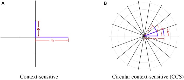
Gliomas have the highest mortality rate and prevalence among the primary brain tumors. In this study, we proposed a supervised brain tumor segmentation method which detects diverse tumoral structures of both high grade gliomas and low grade gliomas in magnetic resonance imaging (MRI) images based on two types of features, the gradient features and the context-sensitive features. Two-dimensional gradient and three-dimensional gradient information was fully utilized to capture the gradient change. Furthermore, we proposed a circular context-sensitive feature which captures context information effectively. These features, totally 62, were compressed and optimized based on an mRMR algorithm, and random forest was used to classify voxels based on the compact feature set. To overcome the class-imbalanced problem of MRI data, our model was trained on a class-balanced region of interest dataset. We evaluated the proposed method based on the 2015 Brain Tumor Segmentation Challenge database, and the experimental results show a competitive performance.

摘要

神经胶质瘤在原发性脑肿瘤中死亡率和发病率最高。在本研究中,我们提出了一种监督式脑肿瘤分割方法,该方法基于梯度特征和上下文敏感特征这两种特征,在磁共振成像(MRI)图像中检测高级别神经胶质瘤和低级别神经胶质瘤的各种肿瘤结构。充分利用二维梯度和三维梯度信息来捕捉梯度变化。此外,我们提出了一种圆形上下文敏感特征,可有效捕捉上下文信息。这些总共62个特征基于mRMR算法进行压缩和优化,并使用随机森林基于紧凑特征集对体素进行分类。为了克服MRI数据的类别不平衡问题,我们的模型在类别平衡的感兴趣区域数据集上进行训练。我们基于2015年脑肿瘤分割挑战赛数据库对所提出的方法进行了评估,实验结果显示出具有竞争力的性能。

https://cdn.ncbi.nlm.nih.gov/pmc/blobs/367e/6427904/d18bbaf6a4fa/fnins-13-00144-g0004.jpg
https://cdn.ncbi.nlm.nih.gov/pmc/blobs/367e/6427904/51ef498faeb5/fnins-13-00144-g0001.jpg
https://cdn.ncbi.nlm.nih.gov/pmc/blobs/367e/6427904/64575472ba4d/fnins-13-00144-g0002.jpg
https://cdn.ncbi.nlm.nih.gov/pmc/blobs/367e/6427904/55b25a8a6e4c/fnins-13-00144-g0003.jpg
https://cdn.ncbi.nlm.nih.gov/pmc/blobs/367e/6427904/d18bbaf6a4fa/fnins-13-00144-g0004.jpg

相似文献

1
Supervised Brain Tumor Segmentation Based on Gradient and Context-Sensitive Features.基于梯度和上下文敏感特征的监督式脑肿瘤分割
Front Neurosci. 2019 Mar 14;13:144. doi: 10.3389/fnins.2019.00144. eCollection 2019.
2
Region-related focal loss for 3D brain tumor MRI segmentation.基于区域的 3D 脑肿瘤 MRI 分割的焦点损失。
Med Phys. 2023 Jul;50(7):4325-4339. doi: 10.1002/mp.16244. Epub 2023 Feb 16.
3
RFDCR: Automated brain lesion segmentation using cascaded random forests with dense conditional random fields.RFDCR:使用级联随机森林和密集条件随机场进行自动脑损伤分割。
Neuroimage. 2020 May 1;211:116620. doi: 10.1016/j.neuroimage.2020.116620. Epub 2020 Feb 11.
4
Machine-Learning-Based Radiomics for Classifying Glioma Grade from Magnetic Resonance Images of the Brain.基于机器学习的脑磁共振图像胶质瘤分级放射组学
J Pers Med. 2023 May 30;13(6):920. doi: 10.3390/jpm13060920.
5
Supervised learning based multimodal MRI brain tumour segmentation using texture features from supervoxels.基于监督学习的利用超体素纹理特征的多模态 MRI 脑肿瘤分割。
Comput Methods Programs Biomed. 2018 Apr;157:69-84. doi: 10.1016/j.cmpb.2018.01.003. Epub 2018 Jan 11.
6
Voxel-based clustered imaging by multiparameter diffusion tensor images for glioma grading.基于体素的多参数弥散张量成像在胶质瘤分级中的聚类成像。
Neuroimage Clin. 2014 Aug 7;5:396-407. doi: 10.1016/j.nicl.2014.08.001. eCollection 2014.
7
Self-Supervised Multi-Modal Hybrid Fusion Network for Brain Tumor Segmentation.基于自监督多模态混合融合网络的脑肿瘤分割。
IEEE J Biomed Health Inform. 2022 Nov;26(11):5310-5320. doi: 10.1109/JBHI.2021.3109301. Epub 2022 Nov 10.
8
Brain tumor segmentation from multimodal magnetic resonance images via sparse representation.基于稀疏表示的多模态磁共振图像脑肿瘤分割
Artif Intell Med. 2016 Oct;73:1-13. doi: 10.1016/j.artmed.2016.08.004. Epub 2016 Sep 6.
9
Detection and Grading of Gliomas Using a Novel Two-Phase Machine Learning Method Based on MRI Images.基于MRI图像的新型两阶段机器学习方法用于胶质瘤的检测与分级
Front Neurosci. 2021 May 14;15:650629. doi: 10.3389/fnins.2021.650629. eCollection 2021.
10
GLISTRboost: Combining Multimodal MRI Segmentation, Registration, and Biophysical Tumor Growth Modeling with Gradient Boosting Machines for Glioma Segmentation.GLISTRboost:结合多模态磁共振成像分割、配准和生物物理肿瘤生长建模与梯度提升机进行胶质瘤分割
Brainlesion. 2016;9556:144-155. doi: 10.1007/978-3-319-30858-6_1.

引用本文的文献

1
Magnetic resonance image-based brain tumour segmentation methods: A systematic review.基于磁共振成像的脑肿瘤分割方法:一项系统综述。
Digit Health. 2022 Mar 16;8:20552076221074122. doi: 10.1177/20552076221074122. eCollection 2022 Jan-Dec.
2
State-of-the-Art Challenges and Perspectives in Multi-Organ Cancer Diagnosis via Deep Learning-Based Methods.基于深度学习方法的多器官癌症诊断的前沿挑战与展望
Cancers (Basel). 2021 Nov 4;13(21):5546. doi: 10.3390/cancers13215546.
3
Computer simulation of tumour resection-induced brain deformation by a meshless approach.

本文引用的文献

1
Developing a Multi-Dose Computational Model for Drug-Induced Hepatotoxicity Prediction Based on Toxicogenomics Data.基于毒理基因组学数据开发用于药物性肝毒性预测的多剂量计算模型。
IEEE/ACM Trans Comput Biol Bioinform. 2019 Jul-Aug;16(4):1231-1239. doi: 10.1109/TCBB.2018.2858756. Epub 2018 Jul 23.
2
A deep learning model integrating FCNNs and CRFs for brain tumor segmentation.基于 FCNNs 和 CRFs 的深度学习模型在脑肿瘤分割中的应用。
Med Image Anal. 2018 Jan;43:98-111. doi: 10.1016/j.media.2017.10.002. Epub 2017 Oct 5.
3
Tumor origin detection with tissue-specific miRNA and DNA methylation markers.
无网格方法模拟肿瘤切除引起的脑形变。
Int J Numer Method Biomed Eng. 2022 Jan;38(1):e3539. doi: 10.1002/cnm.3539. Epub 2021 Oct 24.
4
Enhanced Region Growing for Brain Tumor MR Image Segmentation.用于脑肿瘤磁共振图像分割的增强区域生长法
J Imaging. 2021 Feb 1;7(2):22. doi: 10.3390/jimaging7020022.
5
Data Augmentation for Brain-Tumor Segmentation: A Review.用于脑肿瘤分割的数据增强:综述
Front Comput Neurosci. 2019 Dec 11;13:83. doi: 10.3389/fncom.2019.00083. eCollection 2019.
利用组织特异性 miRNA 和 DNA 甲基化标记物进行肿瘤起源检测。
Bioinformatics. 2018 Feb 1;34(3):398-406. doi: 10.1093/bioinformatics/btx622.
4
Pretata: predicting TATA binding proteins with novel features and dimensionality reduction strategy.Pretata:运用新特征和降维策略预测TATA结合蛋白
BMC Syst Biol. 2016 Dec 23;10(Suppl 4):114. doi: 10.1186/s12918-016-0353-5.
5
DALSA: Domain Adaptation for Supervised Learning From Sparsely Annotated MR Images.DALSA:基于稀疏标注磁共振图像的有监督学习领域自适应方法。
IEEE Trans Med Imaging. 2016 Jan;35(1):184-96. doi: 10.1109/TMI.2015.2463078. Epub 2015 Jul 30.
6
The Multimodal Brain Tumor Image Segmentation Benchmark (BRATS).多模态脑肿瘤图像分割基准(BRATS)。
IEEE Trans Med Imaging. 2015 Oct;34(10):1993-2024. doi: 10.1109/TMI.2014.2377694. Epub 2014 Dec 4.
7
State of the art survey on MRI brain tumor segmentation.磁共振脑肿瘤分割的最新技术综述。
Magn Reson Imaging. 2013 Oct;31(8):1426-38. doi: 10.1016/j.mri.2013.05.002. Epub 2013 Jun 20.
8
A survey of MRI-based medical image analysis for brain tumor studies.基于 MRI 的脑肿瘤研究医学图像分析调查。
Phys Med Biol. 2013 Jul 7;58(13):R97-129. doi: 10.1088/0031-9155/58/13/R97. Epub 2013 Jun 6.
9
Development of image-processing software for automatic segmentation of brain tumors in MR images.用于磁共振图像中脑肿瘤自动分割的图像处理软件的开发。
J Med Phys. 2011 Jul;36(3):147-58. doi: 10.4103/0971-6203.83481.
10
Edge focusing.边缘聚焦。
IEEE Trans Pattern Anal Mach Intell. 1987 Jun;9(6):726-41. doi: 10.1109/tpami.1987.4767980.