Department of Medical Oncology/Hematologic Neoplasia, Dana Farber Cancer Institute, Boston, Massachusetts, United States of America.
PLoS One. 2011;6(9):e25351. doi: 10.1371/journal.pone.0025351. Epub 2011 Sep 28.
Clinical responses achieved with FLT3 kinase inhibitors in acute myeloid leukemia (AML) are typically transient and partial. Thus, there is a need for identification of molecular mechanisms of clinical resistance to these drugs. In response, we characterized MOLM13 AML cell lines made resistant to two structurally-independent FLT3 inhibitors.
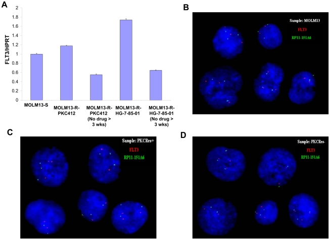
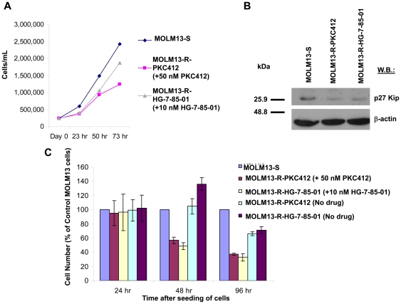
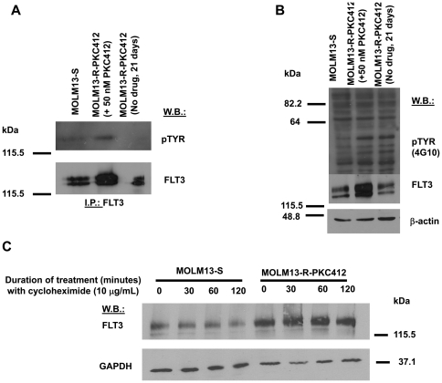
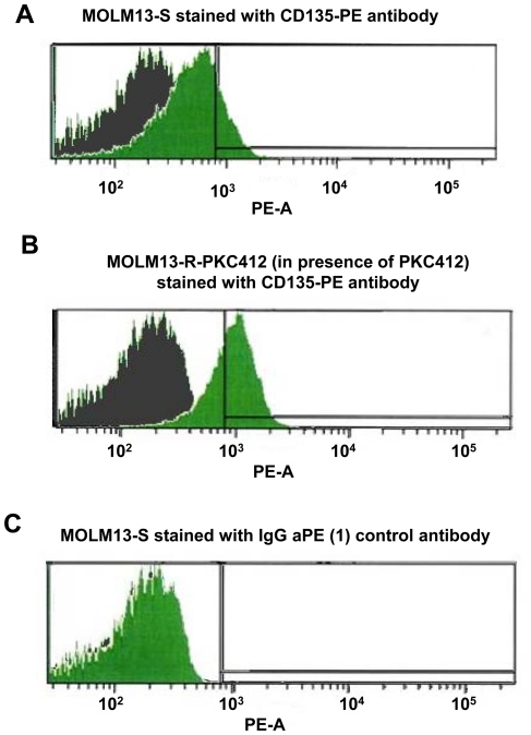
MOLM13 cells were made drug resistant via prolonged exposure to midostaurin and HG-7-85-01, respectively. Cell proliferation was determined by Trypan blue exclusion. Protein expression was assessed by immunoblotting, immunoprecipitation, and flow cytometry. Cycloheximide was used to determine protein half-life. RT-PCR was performed to determine FLT3 mRNA levels, and FISH analysis was performed to determine FLT3 gene expression.
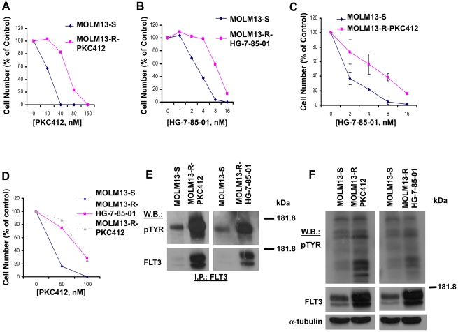
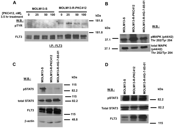
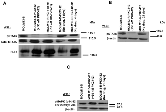
We found that MOLM13 cells readily developed cross-resistance when exposed to either midostaurin or HG-7-85-01. Resistance in both lines was associated with dramatically elevated levels of cell surface FLT3 and elevated levels of phosphor-MAPK, but not phospho-STAT5. The increase in FLT3-ITD expression was at least in part due to reduced turnover of the receptor, with prolonged half-life. Importantly, the drug-resistant phenotype could be rapidly reversed upon withdrawal of either inhibitor. Consistent with this phenotype, no significant evidence of FLT3 gene amplification, kinase domain mutations, or elevated levels of mRNA was observed, suggesting that protein turnover may be part of an auto-regulatory pathway initiated by FLT3 kinase activity. Interestingly, FLT3 inhibitor resistance also correlated with resistance to cytosine arabinoside. Over-expression of FLT3 protein in response to kinase inhibitors may be part of a novel mechanism that could contribute to clinical resistance.
FLT3 激酶抑制剂在急性髓性白血病(AML)中取得的临床应答通常是短暂和部分的。因此,需要确定这些药物临床耐药的分子机制。为此,我们对两种结构上不同的 FLT3 抑制剂耐药的 MOLM13 AML 细胞系进行了特征描述。
通过延长暴露于米哚妥林和 HG-7-85-01 来使 MOLM13 细胞产生耐药性。通过台盼蓝排斥试验测定细胞增殖。通过免疫印迹、免疫沉淀和流式细胞术评估蛋白表达。使用环己酰亚胺测定蛋白半衰期。通过 RT-PCR 测定 FLT3 mRNA 水平,通过 FISH 分析测定 FLT3 基因表达。
我们发现,当暴露于米哚妥林或 HG-7-85-01 时,MOLM13 细胞很容易产生交叉耐药性。两条耐药线均与细胞表面 FLT3 水平显著升高以及 MAPK 磷酸化水平升高相关,但与 STAT5 磷酸化水平升高无关。FLT3-ITD 表达增加至少部分是由于受体周转率降低,半衰期延长。重要的是,在撤回任一抑制剂后,耐药表型可迅速逆转。与该表型一致,未观察到 FLT3 基因扩增、激酶结构域突变或 mRNA 水平升高的显著证据,这表明蛋白周转率可能是由 FLT3 激酶活性启动的自动调节途径的一部分。有趣的是,FLT3 抑制剂耐药性也与阿糖胞苷耐药性相关。对激酶抑制剂的 FLT3 蛋白过表达可能是一种新的机制的一部分,可能有助于临床耐药性。