Departamento de Neurobiología-Investigación, Hospital Ramón y Cajal, IRYCIS, Madrid, Spain.
Mol Neurodegener. 2011 Oct 13;6:72. doi: 10.1186/1750-1326-6-72.
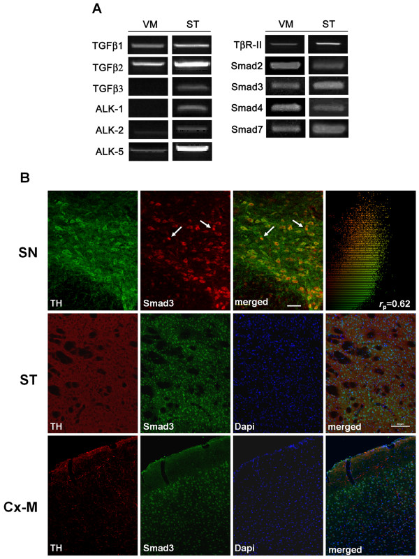
Parkinson's disease (PD) is characterized by dopaminergic neurodegeneration in the substantia nigra (SN). Transforming growth factor-β1 (TGF-β1) levels increase in patients with PD, although the effects of this increment remain unclear. We have examined the mesostriatal system in adult mice deficient in Smad3, a molecule involved in the intracellular TGF-β1 signalling cascade.
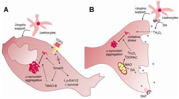
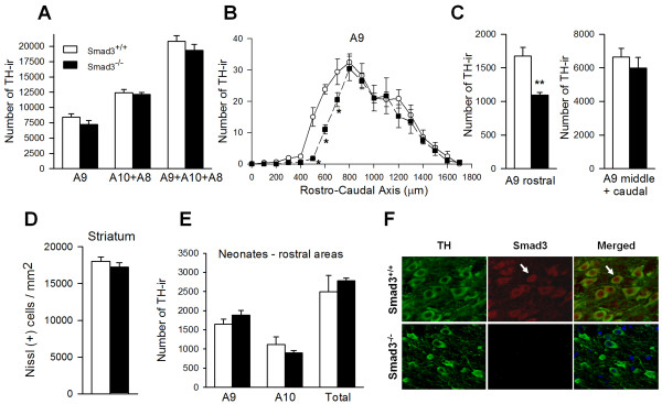
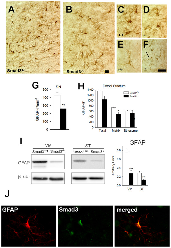
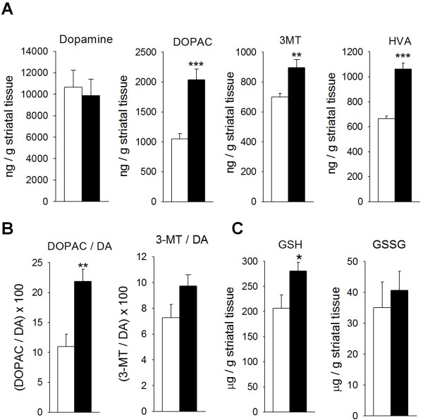
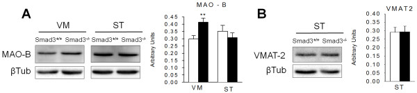
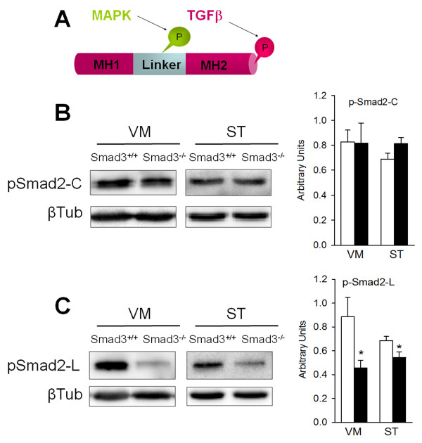
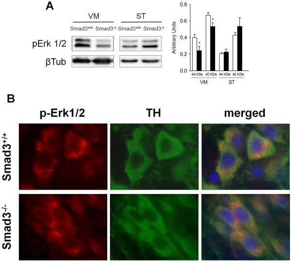
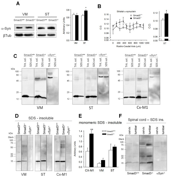
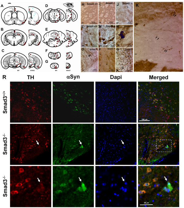
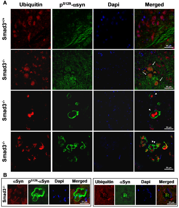
Striatal monoamine oxidase (MAO)-mediated dopamine (DA) catabolism to 3,4-dihydroxyphenylacetic acid (DOPAC) is strongly increased, promoting oxidative stress that is reflected by an increase in glutathione levels. Fewer astrocytes are detected in the ventral midbrain (VM) and striatal matrix, suggesting decreased trophic support to dopaminergic neurons. The SN of these mice has dopaminergic neuronal degeneration in its rostral portion, and the pro-survival Erk1/2 signalling is diminished in nigra dopaminergic neurons, not associated with alterations to p-JNK or p-p38. Furthermore, inclusions of α-synuclein are evident in selected brain areas, both in the perikaryon (SN and paralemniscal nucleus) or neurites (motor and cingulate cortices, striatum and spinal cord). Interestingly, these α-synuclein deposits are detected with ubiquitin and P(S129)-α-synuclein in a core/halo cellular distribution, which resemble those observed in human Lewy bodies (LB).
Smad3 deficiency promotes strong catabolism of DA in the striatum (ST), decrease trophic and astrocytic support to dopaminergic neurons and may induce α-synuclein aggregation, which may be related to early parkinsonism. These data underline a role for Smad3 in α-synuclein and DA homeostasis, and suggest that modulatory molecules of this signalling pathway should be evaluated as possible neuroprotective agents.
帕金森病(PD)的特征是黑质(SN)中的多巴胺能神经退行性变。尽管这种增加的影响尚不清楚,但 PD 患者的转化生长因子-β1(TGF-β1)水平升高。我们检查了成年 Smad3 缺失的小鼠的中脑纹状体系统,Smad3 是参与 TGF-β1 细胞内信号转导级联的分子。
纹状体单胺氧化酶(MAO)介导的多巴胺(DA)代谢为 3,4-二羟基苯乙酸(DOPAC)的速度大大增加,促进了氧化应激,这反映在谷胱甘肽水平的增加上。腹侧中脑(VM)和纹状体基质中的星形胶质细胞减少,表明对多巴胺能神经元的营养支持减少。这些小鼠的 SN 前部有多巴胺能神经元变性,并且 nigra 多巴胺能神经元中的促生存 Erk1/2 信号减弱,与 p-JNK 或 p-p38 的改变无关。此外,α-突触核蛋白的包含物在选定的脑区中是明显的,无论是在胞体(SN 和旁侧楔状核)还是在轴突(运动和扣带皮质、纹状体和脊髓)中。有趣的是,这些α-突触核蛋白沉积物与泛素和 P(S129)-α-突触核蛋白一起在核心/晕细胞分布中被检测到,这类似于在人类路易体(LB)中观察到的分布。
Smad3 缺失促进了纹状体(ST)中 DA 的强烈代谢,减少了对多巴胺能神经元的营养和星形胶质细胞的支持,并且可能诱导α-突触核蛋白聚集,这可能与早期帕金森病有关。这些数据强调了 Smad3 在α-突触核蛋白和 DA 动态平衡中的作用,并表明该信号通路的调节分子应作为潜在的神经保护剂进行评估。