Department of Radiology, University Medical Center Utrecht, PO Box 85500, E01.132, 3508 GA Utrecht, The Netherlands.
Int J Cardiovasc Imaging. 2012 Aug;28(6):1567-75. doi: 10.1007/s10554-011-9954-7. Epub 2011 Oct 15.
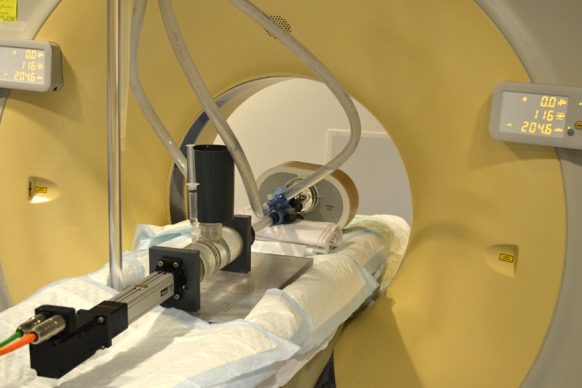
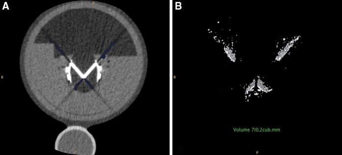
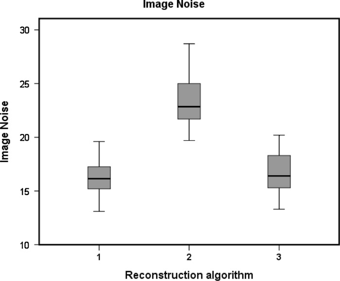
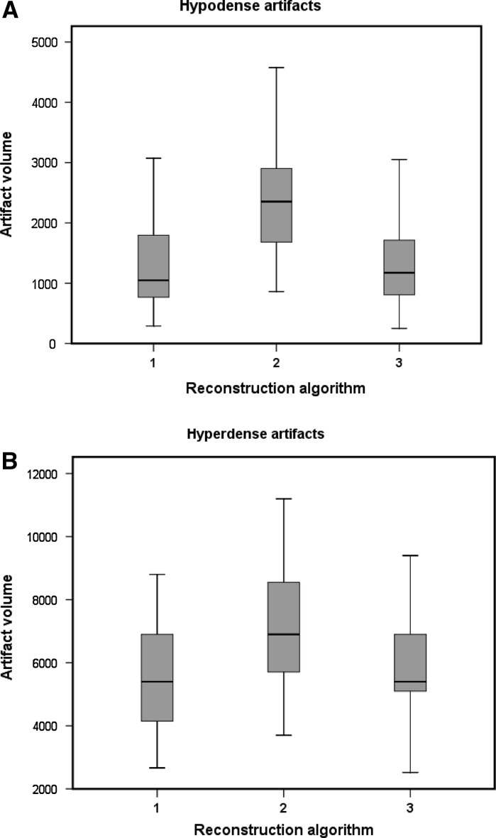
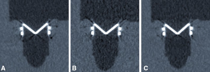
Multidetector-row CT is promising for prosthetic heart valve (PHV) assessment but retrospectively ECG-gated scanning has a considerable radiation dose. Recently introduced iterative reconstruction (IR) algorithms may enable radiation dose reduction with retained image quality. Furthermore, PHV image quality on the CT scan mainly depends on extent of PHV artifacts. IR may decrease streak artifacts. We compared image noise and artifact volumes in scans of mechanical PHVs reconstructed with conventional filtered back projection (FBP) to lower dose scans reconstructed with IR. Four different PHVs (St. Jude, Carbomedics, ON-X and Medtronic Hall) were scanned in a pulsatile in vitro model. Ten retrospectively ECG-gated CT scans were performed of each PHV at 120 kV, 600 mAs (high-dose CTDI(vol) 35.3 mGy) and 120 kV, 300 mAs (low-dose CTDI(vol) 17.7 mGy) on a 64 detector-row scanner. Diastolic and systolic images were reconstructed with FBP (high and low-dose) and the IR algorithm (low-dose only). Hypo- and hyperdense artifact volumes were determined using two threshold filters. Image noise was measured. Mean hypo- and hyperdense artifact volumes (mm(3)) were 1,235/5,346 (high-dose FBP); 2,405/6,877 (low-dose FBP) and 1,218/5,333 (low-dose IR). Low-dose IR reconstructions had similar image noise compared to high-dose FBP (16.5 ± 1.7 vs. 16.3 ± 1.6, mean ± SD, respectively, P = 1.0). IR allows ECG-gated PHV imaging with similar image noise and PHV artifacts at 50% less dose compared to conventional FBP in an pulsatile in vitro model.
多排螺旋 CT 对人工心脏瓣膜(PHV)评估具有很大的应用潜力,但回顾性心电门控扫描的辐射剂量较大。最近引入的迭代重建(IR)算法可以在保持图像质量的同时降低辐射剂量。此外,CT 扫描中 PHV 的图像质量主要取决于 PHV 伪影的程度。IR 可能会减少条纹伪影。我们比较了用传统滤波反投影(FBP)重建的机械 PHV 的低剂量扫描与用 IR 重建的机械 PHV 的图像噪声和伪影体积。在一个脉动的体外模型中,对四种不同的 PHV(St. Jude、Carbomedics、ON-X 和 Medtronic Hall)进行了扫描。对每个 PHV 分别在 120kV、600mA(高剂量 CTDI(vol) 35.3mGy)和 120kV、300mA(低剂量 CTDI(vol) 17.7mGy)进行了 10 次回顾性心电门控 CT 扫描,在一台 64 排探测器的扫描仪上完成。舒张期和收缩期图像分别用 FBP(高剂量和低剂量)和 IR 算法(仅低剂量)重建。使用两个阈值滤波器确定低和高密伪影的体积。测量图像噪声。低剂量 IR 重建的平均低和高密伪影体积(mm(3))分别为 1235/5346(高剂量 FBP)、2405/6877(低剂量 FBP)和 1218/5333(低剂量 IR)。与高剂量 FBP 相比,低剂量 IR 重建具有相似的图像噪声(分别为 16.5±1.7 和 16.3±1.6,平均值±标准差,P=1.0)。在一个脉动的体外模型中,IR 允许在降低 50%的剂量下进行 ECG 门控 PHV 成像,与传统的 FBP 相比,具有相似的图像噪声和 PHV 伪影。