Wolfson Institute for Biomedical Research, University College London, London WCIE 6BT, United Kingdom.
J Biol Chem. 2011 Dec 16;286(50):43172-81. doi: 10.1074/jbc.M111.289777. Epub 2011 Oct 20.
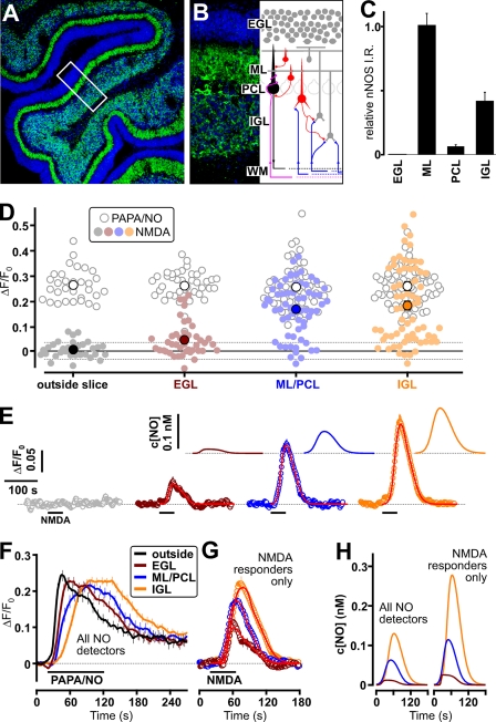
Nitric oxide (NO) is a widespread signaling molecule with potentially multifarious actions of relevance to health and disease. A fundamental determinant of how it acts is its concentration, but there remains a lack of coherent information on the patterns of NO release from its sources, such as neurons or endothelial cells, in either normal or pathological conditions. We have used detector cells having the highest recorded NO sensitivity to monitor NO release from brain tissue quantitatively and in real time. Stimulation of NMDA receptors, which are coupled to activation of neuronal NO synthase, routinely generated NO signals from neurons in cerebellar slices. The average computed peak NO concentrations varied across the anatomical layers of the cerebellum, from 12 to 130 pm. The mean value found in the hippocampus was 200 pm. Much variation in the amplitudes recorded by individual detector cells was observed, this being attributable to their location at variable distances from the NO sources. From fits to the data, the NO concentrations at the source surfaces were 120 pm to 1.4 nm, and the underlying rates of NO generation were 36-350 nm/s, depending on area. Our measurements are 4-5 orders of magnitude lower than reported by some electrode recordings in cerebellum or hippocampus. In return, they establish coherence between the NO concentrations able to elicit physiological responses in target cells through guanylyl cyclase-linked NO receptors, the concentrations that neuronal NO synthase is predicted to generate locally, and the concentrations that neurons actually produce.
一氧化氮(NO)是一种广泛存在的信号分子,具有与健康和疾病相关的潜在多种作用。其作用方式的一个基本决定因素是其浓度,但对于神经元或内皮细胞等来源在正常或病理条件下的 NO 释放模式,仍然缺乏一致的信息。我们使用具有记录到的最高 NO 灵敏度的检测细胞来定量和实时监测脑组织中 NO 的释放。NMDA 受体的刺激与神经元型一氧化氮合酶的激活偶联,通常会从小脑切片中的神经元产生 NO 信号。计算得出的平均峰值 NO 浓度在小脑的解剖层之间变化,从 12 到 130 pm。在海马体中发现的平均值为 200 pm。个体检测细胞记录的幅度变化很大,这归因于它们与 NO 源的距离不同。根据数据拟合,源表面的 NO 浓度为 120 pm 至 1.4nm,基础 NO 生成速率为 36-350nm/s,具体取决于面积。我们的测量值比一些在小脑或海马体中的电极记录报告的值低 4-5 个数量级。作为回报,它们在能够通过与鸟苷酸环化酶偶联的 NO 受体在靶细胞中引发生理反应的 NO 浓度、神经元型一氧化氮合酶局部预测产生的浓度以及神经元实际产生的浓度之间建立了一致性。