• 文献检索
  • 文档翻译
  • 深度研究
  • 学术资讯
  • Suppr Zotero 插件Zotero 插件
  • 邀请有礼
  • 套餐&价格
  • 历史记录
应用&插件
Suppr Zotero 插件Zotero 插件浏览器插件Mac 客户端Windows 客户端微信小程序
定价
高级版会员购买积分包购买API积分包
服务
文献检索文档翻译深度研究API 文档MCP 服务
关于我们
关于 Suppr公司介绍联系我们用户协议隐私条款
关注我们

Suppr 超能文献

核心技术专利:CN118964589B侵权必究
粤ICP备2023148730 号-1Suppr @ 2026

文献检索

告别复杂PubMed语法,用中文像聊天一样搜索,搜遍4000万医学文献。AI智能推荐,让科研检索更轻松。

立即免费搜索

文件翻译

保留排版,准确专业,支持PDF/Word/PPT等文件格式,支持 12+语言互译。

免费翻译文档

深度研究

AI帮你快速写综述,25分钟生成高质量综述,智能提取关键信息,辅助科研写作。

立即免费体验

拉曼光谱法:一种在立体定向乳腺粗针活检过程中识别微钙化的实时工具。

Raman spectroscopy: a real-time tool for identifying microcalcifications during stereotactic breast core needle biopsies.

作者信息

Saha A, Barman I, Dingari N C, McGee S, Volynskaya Z, Galindo L H, Liu W, Plecha D, Klein N, Dasari R R, Fitzmaurice M

出版信息

Biomed Opt Express. 2011 Oct 1;2(10):2792-803. doi: 10.1364/BOE.2.002792. Epub 2011 Sep 14.

DOI:10.1364/BOE.2.002792
PMID:22025985
原文链接:https://pmc.ncbi.nlm.nih.gov/articles/PMC3191446/
Abstract

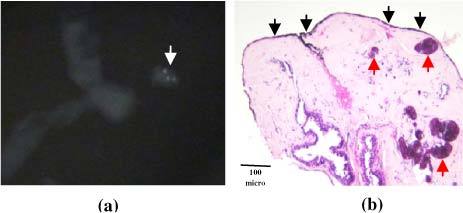
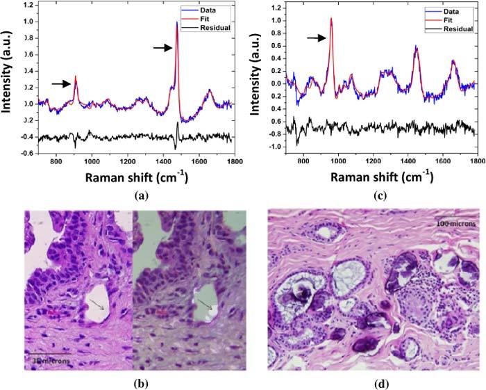
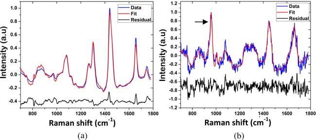
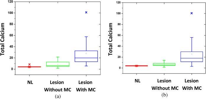
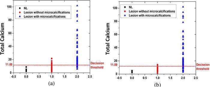
Microcalcifications are an early mammographic sign of breast cancer and a target for stereotactic breast needle biopsy. We present here a Raman spectroscopic tool for detecting microcalcifications in breast tissue based on their chemical composition. We collected ex vivo Raman spectra from 159 tissue sites in fresh stereotactic breast needle biopsies from 33 patients, including 54 normal sites, 75 lesions with microcalcifications and 30 lesions without microcalcifications. Application of our Raman technique resulted in a positive predictive value of 97% for detecting microcalcifications. This study shows that Raman spectroscopy has the potential to detect microcalcifications during stereotactic breast core biopsies and provide real-time feedback to radiologists, thus reducing non-diagnostic and false negative biopsies.

摘要

微钙化是乳腺癌的早期乳腺X线征象,也是立体定向乳腺针吸活检的目标。我们在此展示一种基于化学成分检测乳腺组织中微钙化的拉曼光谱工具。我们从33例患者的新鲜立体定向乳腺针吸活检的159个组织部位收集了离体拉曼光谱,包括54个正常部位、75个有微钙化的病变部位和30个无微钙化的病变部位。我们的拉曼技术应用于检测微钙化的阳性预测值为97%。这项研究表明,拉曼光谱有潜力在立体定向乳腺粗针活检过程中检测微钙化,并向放射科医生提供实时反馈,从而减少非诊断性和假阴性活检。

https://cdn.ncbi.nlm.nih.gov/pmc/blobs/ba7b/3191446/3e13cb4638a9/boe-2-10-2792-g006.jpg
https://cdn.ncbi.nlm.nih.gov/pmc/blobs/ba7b/3191446/c8c2fea10235/boe-2-10-2792-g001.jpg
https://cdn.ncbi.nlm.nih.gov/pmc/blobs/ba7b/3191446/f820d44e0da7/boe-2-10-2792-g002.jpg
https://cdn.ncbi.nlm.nih.gov/pmc/blobs/ba7b/3191446/bcaf4a559d54/boe-2-10-2792-g003.jpg
https://cdn.ncbi.nlm.nih.gov/pmc/blobs/ba7b/3191446/172e3ee6b8f1/boe-2-10-2792-g004.jpg
https://cdn.ncbi.nlm.nih.gov/pmc/blobs/ba7b/3191446/7ed228d24c06/boe-2-10-2792-g005.jpg
https://cdn.ncbi.nlm.nih.gov/pmc/blobs/ba7b/3191446/3e13cb4638a9/boe-2-10-2792-g006.jpg

相似文献

1
Raman spectroscopy: a real-time tool for identifying microcalcifications during stereotactic breast core needle biopsies.拉曼光谱法:一种在立体定向乳腺粗针活检过程中识别微钙化的实时工具。
Biomed Opt Express. 2011 Oct 1;2(10):2792-803. doi: 10.1364/BOE.2.002792. Epub 2011 Sep 14.
2
Application of Raman spectroscopy to identify microcalcifications and underlying breast lesions at stereotactic core needle biopsy.拉曼光谱在立体定向核心针活检中识别微钙化及潜在乳腺病变中的应用。
Cancer Res. 2013 Jun 1;73(11):3206-15. doi: 10.1158/0008-5472.CAN-12-2313.
3
Precision of Raman spectroscopy measurements in detection of microcalcifications in breast needle biopsies.拉曼光谱测量在乳腺针吸活检中检测微钙化的精确性。
Anal Chem. 2012 Aug 7;84(15):6715-22. doi: 10.1021/ac3011439. Epub 2012 Jul 12.
4
Diagnostic power of diffuse reflectance spectroscopy for targeted detection of breast lesions with microcalcifications.弥散反射光谱对伴微钙化的乳腺病灶进行靶向检测的诊断效能。
Proc Natl Acad Sci U S A. 2013 Jan 8;110(2):471-6. doi: 10.1073/pnas.1215473110. Epub 2012 Dec 24.
5
Development and comparative assessment of Raman spectroscopic classification algorithms for lesion discrimination in stereotactic breast biopsies with microcalcifications.发展和比较评估用于立体定向乳腺活检中微钙化病变鉴别拉曼光谱分类算法。
J Biophotonics. 2013 Apr;6(4):371-81. doi: 10.1002/jbio.201200098. Epub 2012 Jul 20.
6
Raman spectroscopic sensing of carbonate intercalation in breast microcalcifications at stereotactic biopsy.立体定向活检时乳腺微钙化中碳酸盐嵌入的拉曼光谱传感
Sci Rep. 2015 Apr 30;5:9907. doi: 10.1038/srep09907.
7
Raman Spectroscopy Reveals That Biochemical Composition of Breast Microcalcifications Correlates with Histopathologic Features.拉曼光谱分析显示乳腺微钙化的生化成分与组织病理学特征相关。
Cancer Res. 2020 Apr 15;80(8):1762-1772. doi: 10.1158/0008-5472.CAN-19-3204. Epub 2020 Feb 24.
8
Automated stereotactic core needle biopsy of microcalcifications with correlation to surgical biopsy.微钙化灶的自动立体定向芯针活检及其与手术活检的相关性
Acta Radiol. 1999 Jul;40(4):390-3. doi: 10.3109/02841859909177752.
9
Add-on device for stereotactic core-needle breast biopsy: how many biopsy specimens are needed for a reliable diagnosis?立体定向芯针乳腺活检的附加装置:进行可靠诊断需要多少活检样本?
Radiology. 2005 Sep;236(3):801-9. doi: 10.1148/radiol.2363040782. Epub 2005 Jul 14.
10
Learning curve for add-on stereotactic core needle breast biopsy.附加立体定向芯针乳腺活检的学习曲线
Acta Radiol. 2006 Jun;47(5):454-60. doi: 10.1080/02841850600702168.

引用本文的文献

1
Exploration of utility of combined optical photothermal infrared and Raman imaging for investigating the chemical composition of microcalcifications in breast cancer.联合光学光热红外和拉曼成像在研究乳腺癌微钙化化学成分中的应用探讨。
Anal Methods. 2023 Mar 30;15(13):1620-1630. doi: 10.1039/d2ay01197b.
2
Raman Spectroscopy: A Personalized Decision-Making Tool on Clinicians' Hands for In Situ Cancer Diagnosis and Surgery Guidance.拉曼光谱:临床医生手中用于原位癌症诊断和手术指导的个性化决策工具。
Cancers (Basel). 2022 Feb 23;14(5):1144. doi: 10.3390/cancers14051144.
3
Breast microcalcifications: Past, present and future (Review).

本文引用的文献

1
Wavelength selection-based nonlinear calibration for transcutaneous blood glucose sensing using Raman spectroscopy.基于波长选择的拉曼光谱无创血糖检测中的非线性校准。
J Biomed Opt. 2011 Aug;16(8):087009. doi: 10.1117/1.3611006.
2
New relationships between breast microcalcifications and cancer.乳腺微钙化与癌症的新关系。
Br J Cancer. 2010 Sep 28;103(7):1034-9. doi: 10.1038/sj.bjc.6605873. Epub 2010 Sep 14.
3
Diagnosing breast cancer using Raman spectroscopy: prospective analysis.使用拉曼光谱诊断乳腺癌:前瞻性分析。
乳腺微钙化:过去、现在与未来(综述)
Mol Clin Oncol. 2022 Apr;16(4):81. doi: 10.3892/mco.2022.2514. Epub 2022 Feb 10.
4
Raman spectroscopy: current applications in breast cancer diagnosis, challenges and future prospects.拉曼光谱:在乳腺癌诊断中的当前应用、挑战及未来前景
Br J Cancer. 2022 May;126(8):1125-1139. doi: 10.1038/s41416-021-01659-5. Epub 2021 Dec 10.
5
A New Look into Cancer-A Review on the Contribution of Vibrational Spectroscopy on Early Diagnosis and Surgery Guidance.癌症新视角——振动光谱在早期诊断和手术指导中的作用综述
Cancers (Basel). 2021 Oct 24;13(21):5336. doi: 10.3390/cancers13215336.
6
Quantitative chemical imaging of breast calcifications in association with neoplastic processes.定量化学成像在乳腺钙化与肿瘤过程中的相关性研究。
Theranostics. 2020 Apr 27;10(13):5865-5878. doi: 10.7150/thno.43325. eCollection 2020.
7
Optical Imaging Approaches to Investigating Radiation Resistance.用于研究辐射抗性的光学成像方法。
Front Oncol. 2019 Nov 5;9:1152. doi: 10.3389/fonc.2019.01152. eCollection 2019.
8
Applications of Raman spectroscopy in cancer diagnosis.拉曼光谱在癌症诊断中的应用。
Cancer Metastasis Rev. 2018 Dec;37(4):691-717. doi: 10.1007/s10555-018-9770-9.
9
Relationships between pathology and crystal structure in breast calcifications: an X-ray diffraction study in histological sections.乳腺钙化灶的病理学与晶体结构之间的关系:组织切片的X射线衍射研究
NPJ Breast Cancer. 2016 Sep 14;2:16029. doi: 10.1038/npjbcancer.2016.29. eCollection 2016.
10
Label-Free Raman Imaging to Monitor Breast Tumor Signatures.无标记拉曼成像监测乳腺癌标志物。
Technol Cancer Res Treat. 2017 Aug;16(4):461-469. doi: 10.1177/1533034616655953. Epub 2016 Jul 4.
J Biomed Opt. 2009 Sep-Oct;14(5):054023. doi: 10.1117/1.3247154.
4
Diagnosing breast cancer using diffuse reflectance spectroscopy and intrinsic fluorescence spectroscopy.使用漫反射光谱和固有荧光光谱诊断乳腺癌。
J Biomed Opt. 2008 Mar-Apr;13(2):024012. doi: 10.1117/1.2909672.
5
Trends in breast cancer screening and diagnosis.乳腺癌筛查与诊断的趋势
Cleve Clin J Med. 2008 Mar;75 Suppl 1:S2-9. doi: 10.3949/ccjm.75.suppl_1.s2.
6
Use of near-infrared Raman spectroscopy for identification of atherosclerotic plaques in the carotid artery.利用近红外拉曼光谱法识别颈动脉粥样硬化斑块。
Photomed Laser Surg. 2007 Dec;25(6):482-6. doi: 10.1089/pho.2007.2111.
7
Identification of calcifications in cardiac valves by near infrared Raman spectroscopy.通过近红外拉曼光谱法识别心脏瓣膜中的钙化
Photomed Laser Surg. 2007 Aug;25(4):287-90. doi: 10.1089/pho.2007.2100.
8
Subsurface probing of calcifications with spatially offset Raman spectroscopy (SORS): future possibilities for the diagnosis of breast cancer.利用空间偏移拉曼光谱法(SORS)对钙化灶进行皮下探测:乳腺癌诊断的未来可能性。
Analyst. 2007 Sep;132(9):899-905. doi: 10.1039/b705029a. Epub 2007 Jul 2.
9
Prospects for the diagnosis of breast cancer by noninvasive probing of calcifications using transmission Raman spectroscopy.利用透射拉曼光谱对钙化灶进行无创探测诊断乳腺癌的前景。
J Biomed Opt. 2007 Mar-Apr;12(2):024008. doi: 10.1117/1.2718934.
10
Chemical concentration measurement in blood serum and urine samples using liquid-core optical fiber Raman spectroscopy.利用液芯光纤拉曼光谱法测量血清和尿液样本中的化学物质浓度。
Appl Opt. 2007 Apr 1;46(10):1726-34. doi: 10.1364/ao.46.001726.