• 文献检索
  • 文档翻译
  • 深度研究
  • 学术资讯
  • Suppr Zotero 插件Zotero 插件
  • 邀请有礼
  • 套餐&价格
  • 历史记录
应用&插件
Suppr Zotero 插件Zotero 插件浏览器插件Mac 客户端Windows 客户端微信小程序
定价
高级版会员购买积分包购买API积分包
服务
文献检索文档翻译深度研究API 文档MCP 服务
关于我们
关于 Suppr公司介绍联系我们用户协议隐私条款
关注我们

Suppr 超能文献

核心技术专利:CN118964589B侵权必究
粤ICP备2023148730 号-1Suppr @ 2026

文献检索

告别复杂PubMed语法,用中文像聊天一样搜索,搜遍4000万医学文献。AI智能推荐,让科研检索更轻松。

立即免费搜索

文件翻译

保留排版,准确专业,支持PDF/Word/PPT等文件格式,支持 12+语言互译。

免费翻译文档

深度研究

AI帮你快速写综述,25分钟生成高质量综述,智能提取关键信息,辅助科研写作。

立即免费体验

用于口腔锥形束 CT 的质量控制体模的研制与适用性。

Development and applicability of a quality control phantom for dental cone-beam CT.

机构信息

Oral Imaging Center, Faculty of Medicine, Katholieke Universiteit Leuven, Leuven, Belgium.

出版信息

J Appl Clin Med Phys. 2011 Nov 15;12(4):3478. doi: 10.1120/jacmp.v12i4.3478.

DOI:10.1120/jacmp.v12i4.3478
PMID:22089004
原文链接:https://pmc.ncbi.nlm.nih.gov/articles/PMC5718749/
Abstract

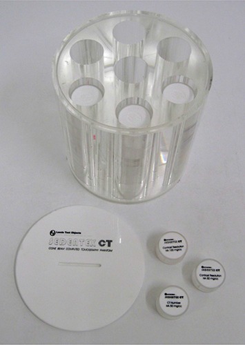
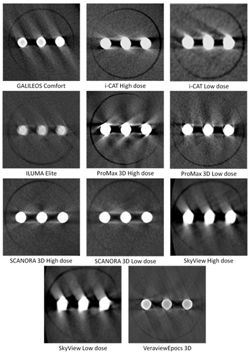
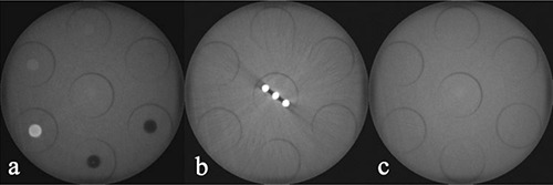
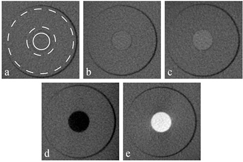
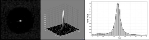
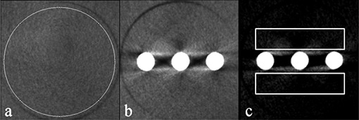
Cone-beam CT (CBCT) has shown to be a useful imaging modality for various dentomaxillofacial applications. However, optimization and quality control of dental CBCT devices is hampered due to the lack of an appropriate tool for image quality assessment. To investigate the application of different image quality parameters for CBCT, a prototype polymethyl methacrylate (PMMA) cylindrical phantom with inserts for image quality analysis was developed. Applicability and reproducibility of the phantom were assessed using seven CBCT devices with different scanning protocols. Image quality parameters evaluated were: CT number correlation, contrast resolution, image homogeneity and uniformity, point spread function, and metal artifacts. Deviations of repeated measurements were between 0.0% and 3.3%. Correlation coefficients of CBCT voxel values with CT numbers ranged between 0.68 and 1.00. Contrast-to-noise ratio (CNR) values were much lower for hydroxyapatite (0 < CNR < 7.7) than for air and aluminum (5.0 < CNR < 32.8). Noise values ranged between 35 and 419. The uniformity index was between 3.3% and 11.9%. Full width at half maximum (FWHM) measurements varied between 0.43 mm and 1.07 mm. The increase of mean voxel values surrounding metal objects ranged between 6.7% and 43.0%. Results from preliminary analyses of the prototype quality control phantom showed its potential for routine quality assurance on CBCT. Large differences in image quality performance were seen between CBCT devices. Based on the initial evaluations, the phantom can be optimized and validated.

摘要

锥形束 CT(CBCT)已被证明是一种用于各种口腔颌面应用的有用成像方式。然而,由于缺乏用于图像质量评估的适当工具,牙科 CBCT 设备的优化和质量控制受到阻碍。为了研究不同图像质量参数在 CBCT 中的应用,开发了一种带有用于图像质量分析插件的原型聚甲基丙烯酸甲酯(PMMA)圆柱形体模。使用具有不同扫描协议的七种 CBCT 设备评估了体模的适用性和可重复性。评估的图像质量参数包括:CT 值相关性、对比度分辨率、图像均匀性和一致性、点扩散函数和金属伪影。重复测量的偏差在 0.0%到 3.3%之间。CBCT 体素值与 CT 值的相关系数在 0.68 到 1.00 之间。羟磷灰石的对比噪声比(CNR)值要低得多(0 < CNR < 7.7),而空气和铝的 CNR 值要高得多(5.0 < CNR < 32.8)。噪声值在 35 到 419 之间。均匀性指数在 3.3%到 11.9%之间。半最大值全宽(FWHM)测量值在 0.43 毫米到 1.07 毫米之间。金属物体周围平均体素值的增加幅度在 6.7%到 43.0%之间。原型质量控制体模初步分析结果表明,它具有在 CBCT 上进行常规质量保证的潜力。不同的 CBCT 设备之间的图像质量性能存在很大差异。基于初始评估,体模可以进行优化和验证。

https://cdn.ncbi.nlm.nih.gov/pmc/blobs/53f7/5718749/007e54021e1f/ACM2-12-245-g006.jpg
https://cdn.ncbi.nlm.nih.gov/pmc/blobs/53f7/5718749/b2d197eba4f6/ACM2-12-245-g001.jpg
https://cdn.ncbi.nlm.nih.gov/pmc/blobs/53f7/5718749/2349e6bd33c9/ACM2-12-245-g002.jpg
https://cdn.ncbi.nlm.nih.gov/pmc/blobs/53f7/5718749/dedae04834c4/ACM2-12-245-g003.jpg
https://cdn.ncbi.nlm.nih.gov/pmc/blobs/53f7/5718749/389d62ea15d9/ACM2-12-245-g004.jpg
https://cdn.ncbi.nlm.nih.gov/pmc/blobs/53f7/5718749/bd30ae9d1ebd/ACM2-12-245-g005.jpg
https://cdn.ncbi.nlm.nih.gov/pmc/blobs/53f7/5718749/007e54021e1f/ACM2-12-245-g006.jpg

相似文献

1
Development and applicability of a quality control phantom for dental cone-beam CT.用于口腔锥形束 CT 的质量控制体模的研制与适用性。
J Appl Clin Med Phys. 2011 Nov 15;12(4):3478. doi: 10.1120/jacmp.v12i4.3478.
2
Cone-beam CT dose and imaging performance evaluation with a modular, multipurpose phantom.使用模块化、多用途体模评估锥形束 CT 剂量和成像性能。
Med Phys. 2020 Feb;47(2):467-479. doi: 10.1002/mp.13952. Epub 2019 Dec 25.
3
Image quality assessment of three cone beam CT machines using the SEDENTEXCT CT phantom.使用 SEDENTEXCT CT 体模评估三种锥形束 CT 机的图像质量。
Dentomaxillofac Radiol. 2013;42(8):20120445. doi: 10.1259/dmfr.20120445.
4
A quality assurance framework for the fully automated and objective evaluation of image quality in cone-beam computed tomography.用于锥形束计算机断层扫描图像质量全自动客观评估的质量保证框架。
Med Phys. 2014 Mar;41(3):031901. doi: 10.1118/1.4863507.
5
Optimization of dental CBCT exposures through mAs reduction.通过降低管电流(mAs)优化牙科锥形束计算机断层扫描(CBCT)曝光。
Dentomaxillofac Radiol. 2015;44(9):20150108. doi: 10.1259/dmfr.20150108. Epub 2015 Jun 19.
6
A pragmatic approach to determine the optimal kVp in cone beam CT: balancing contrast-to-noise ratio and radiation dose.一种确定锥形束CT中最佳千伏峰值的实用方法:平衡对比噪声比与辐射剂量。
Dentomaxillofac Radiol. 2014;43(5):20140059. doi: 10.1259/dmfr.20140059. Epub 2014 Apr 8.
7
Improved image quality of cone beam CT scans for radiotherapy image guidance using fiber-interspaced antiscatter grid.使用纤维间隔反散射网格提高用于放射治疗图像引导的锥形束CT扫描的图像质量。
Med Phys. 2014 Jun;41(6):061910. doi: 10.1118/1.4875978.
8
A method to correct the influence of carbon fiber couchtop and patient positioning device on image quality of cone beam CT.一种校正锥形束 CT 中碳纤维床面和患者定位装置对图像质量影响的方法。
Med Phys. 2010 Jun;37(6):2466-72. doi: 10.1118/1.3425999.
9
A simplified low-cost phantom for image quality assessment of dental cone beam computed tomography unit.一种用于评估口腔锥形束计算机断层摄影设备图像质量的简化经济型体模。
J Med Radiat Sci. 2024 Mar;71(1):78-84. doi: 10.1002/jmrs.738. Epub 2023 Nov 15.
10
Dual-energy imaging method to improve the image quality and the accuracy of dose calculation for cone-beam computed tomography.用于提高锥束计算机断层扫描图像质量和剂量计算准确性的双能成像方法。
Phys Med. 2017 Apr;36:110-118. doi: 10.1016/j.ejmp.2017.03.023. Epub 2017 Apr 4.

引用本文的文献

1
The influence of the field of view and voxel size on the contrast-to-noise ratio in cone-beam computed tomography imaging.视野和体素大小对锥束计算机断层扫描成像中对比度噪声比的影响。
Imaging Sci Dent. 2024 Dec;54(4):362-369. doi: 10.5624/isd.20240096. Epub 2024 Oct 23.
2
Optimization of Fast Non-Local Means Noise Reduction Algorithm Parameter in Computed Tomographic Phantom Images Using 3D Printing Technology.利用3D打印技术对计算机断层扫描体模图像中快速非局部均值降噪算法参数进行优化
Diagnostics (Basel). 2024 Jul 23;14(15):1589. doi: 10.3390/diagnostics14151589.
3
Assessment of cone-beam CT technical image quality indicators and radiation dose for optimal STL model used in visual surgical planning.

本文引用的文献

1
Quality control and patient dosimetry in dental cone beam CT.牙科锥形束CT中的质量控制与患者剂量测定
Radiat Prot Dosimetry. 2010 Apr-May;139(1-3):310-2. doi: 10.1093/rpd/ncq011.
2
Modulation transfer function evaluation of cone beam computed tomography for dental use with the oversampling method.采用过采样方法评估口腔锥形束 CT 的调制传递函数。
Dentomaxillofac Radiol. 2010 Jan;39(1):28-32. doi: 10.1259/dmfr/27069629.
3
On cone-beam computed tomography artifacts induced by titanium implants.基于锥形束 CT 成像的钛种植体伪影研究。
评估锥形束 CT 技术图像质量指标和辐射剂量,以获得用于视觉手术规划的最佳 STL 模型。
Dentomaxillofac Radiol. 2024 Sep 1;53(6):423-433. doi: 10.1093/dmfr/twae026.
4
Feasibility of frozen soft tissues to simulate fresh soft tissue conditions in cone beam CT scans.冷冻软组织在锥形束 CT 扫描中模拟新鲜软组织条件的可行性。
Dentomaxillofac Radiol. 2024 Mar 25;53(3):196-202. doi: 10.1093/dmfr/twae004.
5
A simplified low-cost phantom for image quality assessment of dental cone beam computed tomography unit.一种用于评估口腔锥形束计算机断层摄影设备图像质量的简化经济型体模。
J Med Radiat Sci. 2024 Mar;71(1):78-84. doi: 10.1002/jmrs.738. Epub 2023 Nov 15.
6
Validation of bone mineral density measurement using quantitative CBCT image based on deep learning.基于深度学习的定量 CBCT 图像骨密度测量验证。
Sci Rep. 2023 Jul 24;13(1):11921. doi: 10.1038/s41598-023-38943-8.
7
As low as diagnostically acceptable dose imaging in maxillofacial trauma: a reference quality approach.以可诊断接受剂量行颌面部创伤影像学检查:参考质量方法。
Dentomaxillofac Radiol. 2023 Feb;52(3):20220387. doi: 10.1259/dmfr.20220387. Epub 2023 Jan 23.
8
Impact of metal artefacts on subjective perception of image quality of 13 CBCT devices.金属伪影对 13 种 CBCT 设备图像质量主观感知的影响。
Clin Oral Investig. 2022 Jun;26(6):4457-4466. doi: 10.1007/s00784-022-04409-w. Epub 2022 Feb 15.
9
Diagnostic Performance of Conebeam CT Pixel Values in Active Fenestral Otosclerosis.锥形束CT像素值在活动性镫骨足板耳硬化症中的诊断效能
AJNR Am J Neuroradiol. 2021 Sep;42(9):1667-1670. doi: 10.3174/ajnr.A7192. Epub 2021 Jun 17.
10
An anthropomorphic maxillofacial phantom using 3-dimensional printing, polyurethane rubber and epoxy resin for dental imaging and dosimetry.一种采用 3D 打印、聚氨酯橡胶和环氧树脂制作的拟人化颌面体模,用于口腔成像和剂量学。
Dentomaxillofac Radiol. 2022 Jan 1;51(1):20200323. doi: 10.1259/dmfr.20200323. Epub 2021 Jun 16.
Clin Oral Implants Res. 2010 Jan;21(1):100-7. doi: 10.1111/j.1600-0501.2009.01817.x. Epub 2009 Oct 21.
4
[Quality control of a kV cone beam computed tomography imaging system].[千伏锥形束计算机断层扫描成像系统的质量控制]
Cancer Radiother. 2009 Sep;13(5):345-52. doi: 10.1016/j.canrad.2009.05.009. Epub 2009 Jul 16.
5
Basic principles for use of dental cone beam computed tomography: consensus guidelines of the European Academy of Dental and Maxillofacial Radiology.牙科锥形束计算机断层扫描使用的基本原则:欧洲口腔颌面放射学会共识指南
Dentomaxillofac Radiol. 2009 May;38(4):187-95. doi: 10.1259/dmfr/74941012.
6
Statistical characteristics of streak artifacts on CT images: relationship between streak artifacts and mA s values.CT图像上条纹伪影的统计特征:条纹伪影与毫安秒值之间的关系。
Med Phys. 2009 Feb;36(2):492-9. doi: 10.1118/1.3056554.
7
Evaluation of voxel values in mandibular cancellous bone: relationship between cone-beam computed tomography and multislice helical computed tomography.下颌骨松质骨体素值评估:锥形束计算机断层扫描与多层螺旋计算机断层扫描之间的关系
Clin Oral Implants Res. 2009 May;20(5):503-6. doi: 10.1111/j.1600-0501.2008.01672.x. Epub 2009 Feb 25.
8
Study of the scan uniformity from an i-CAT cone beam computed tomography dental imaging system.i-CAT锥形束计算机断层扫描牙科成像系统扫描均匀性研究
Dentomaxillofac Radiol. 2008 Oct;37(7):365-74. doi: 10.1259/dmfr/13227258.
9
What is cone-beam CT and how does it work?什么是锥形束CT,它是如何工作的?
Dent Clin North Am. 2008 Oct;52(4):707-30, v. doi: 10.1016/j.cden.2008.05.005.
10
Image quality vs radiation dose of four cone beam computed tomography scanners.四台锥形束计算机断层扫描设备的图像质量与辐射剂量对比
Dentomaxillofac Radiol. 2008 Sep;37(6):309-18. doi: 10.1259/dmfr/16770531.