Department of Oral and Maxillofacial Radiology, School of Dentistry, Seoul National University, Seoul, South Korea.
Department of Biomedical Radiation Sciences, Graduate School of Convergence Science and Technology, Seoul National University, Seoul, South Korea.
Sci Rep. 2023 Jul 24;13(1):11921. doi: 10.1038/s41598-023-38943-8.
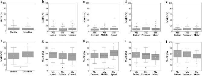
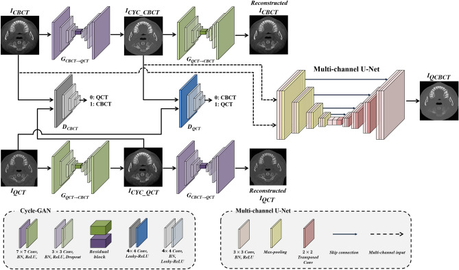
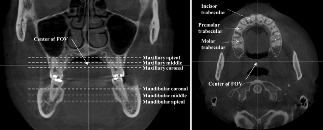
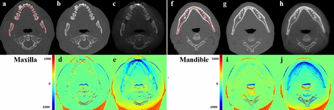
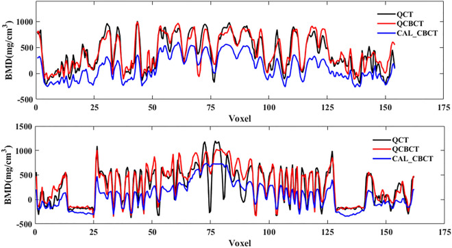
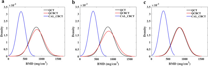
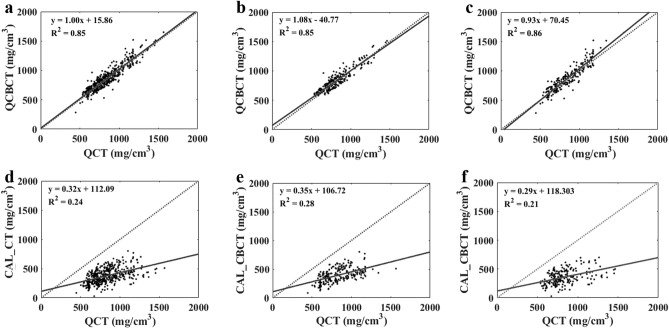
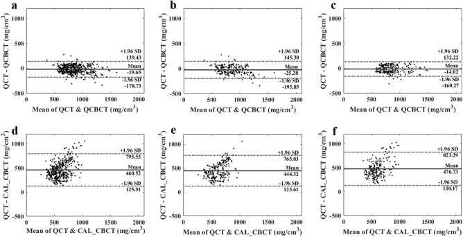
The bone mineral density (BMD) measurement is a direct method of estimating human bone mass for diagnosing osteoporosis, and performed to objectively evaluate bone quality before implant surgery in dental clinics. The objective of this study was to validate the accuracy and reliability of BMD measurements made using quantitative cone-beam CT (CBCT) image based on deep learning by applying the method to clinical data from actual patients. Datasets containing 7500 pairs of CT and CBCT axial slice images from 30 patients were used to train a previously developed deep-learning model (QCBCT-NET). We selected 36 volumes of interest in the CBCT images for each patient in the bone regions of potential implants sites on the maxilla and mandible. We compared the BMDs shown in the quantitative CBCT (QCBCT) images with those in the conventional CBCT (CAL_CBCT) images at the various bone sites of interest across the entire field of view (FOV) using the performance metrics of the MAE, RMSE, MAPE (mean absolute percentage error), R (coefficient of determination), and SEE (standard error of estimation). Compared with the ground truth (QCT) images, the accuracy of the BMD measurements from the QCBCT images showed an RMSE of 83.41 mg/cm, MAE of 67.94 mg/cm, and MAPE of 8.32% across all the bone sites of interest, whereas for the CAL_CBCT images, those values were 491.15 mg/cm, 460.52 mg/cm, and 54.29%, respectively. The linear regression between the QCBCT and QCT images showed a slope of 1.00 and a R of 0.85, whereas for the CAL_CBCT images, those values were 0.32 and 0.24, respectively. The overall SEE between the QCBCT images and QCT images was 81.06 mg/cm, whereas the SEE for the CAL_CBCT images was 109.32 mg/cm. The QCBCT images thus showed better accuracy, linearity, and uniformity than the CAL_CBCT images across the entire FOV. The BMD measurements from the quantitative CBCT images showed high accuracy, linearity, and uniformity regardless of the relative geometric positions of the bone in the potential implant site. When applied to actual patient CBCT images, the CBCT-based quantitative BMD measurement based on deep learning demonstrated high accuracy and reliability across the entire FOV.
骨密度(BMD)测量是一种直接估计人体骨量的方法,用于诊断骨质疏松症,并在牙科诊所进行植体手术前客观评估骨质量。本研究的目的是通过将该方法应用于实际患者的临床数据,验证基于深度学习的定量锥形束 CT(CBCT)图像的 BMD 测量的准确性和可靠性。研究使用了包含 30 名患者的 7500 对 CT 和 CBCT 轴位切片图像的数据集来训练先前开发的深度学习模型(QCBCT-NET)。我们为每个患者在 CBCT 图像中的感兴趣区选择了 36 个感兴趣区,这些患者位于上颌骨和下颌骨的潜在植入部位的骨区。我们使用 MAE、RMSE、MAPE(平均绝对百分比误差)、R(决定系数)和 SEE(估计的标准误差)等性能指标,比较了整个视野(FOV)内各感兴趣区的定量 CBCT(QCBCT)图像与常规 CBCT(CAL_CBCT)图像的 BMD。与地面真实值(QCT)图像相比,QCBCT 图像的 BMD 测量值的准确性在所有感兴趣的骨区显示出 RMSE 为 83.41 mg/cm、MAE 为 67.94 mg/cm 和 MAPE 为 8.32%,而对于 CAL_CBCT 图像,这些值分别为 491.15 mg/cm、460.52 mg/cm 和 54.29%。QCBCT 与 QCT 图像之间的线性回归显示斜率为 1.00,R 为 0.85,而对于 CAL_CBCT 图像,这些值分别为 0.32 和 0.24。QCBCT 图像与 QCT 图像之间的总体 SEE 为 81.06 mg/cm,而 CAL_CBCT 图像的 SEE 为 109.32 mg/cm。在整个 FOV 内,QCBCT 图像的准确性、线性度和均匀性均优于 CAL_CBCT 图像。定量 CBCT 图像的 BMD 测量值无论潜在植入部位骨的相对几何位置如何,均具有较高的准确性、线性度和均匀性。当应用于实际患者的 CBCT 图像时,基于深度学习的基于 CBCT 的定量 BMD 测量在整个 FOV 内表现出较高的准确性和可靠性。