Department of Clinical Biochemistry, Aarhus University Hospital, Norrebrogade 44, Aarhus C, Denmark.
Br J Cancer. 2011 Dec 6;105(12):1850-5. doi: 10.1038/bjc.2011.493. Epub 2011 Nov 17.
We have previously developed (11)C-erlotinib as a new positron emission tomography (PET) tracer and shown that it accumulates in epidermal growth factor receptor (EGFR)-positive lung cancer xenografts in mice. Here, we present a study in patients with non-small cell lung cancer (NSCLC) investigating the feasibility of (11)C-erlotinib PET as a potential method for the identification of lung tumours accumulating erlotinib.
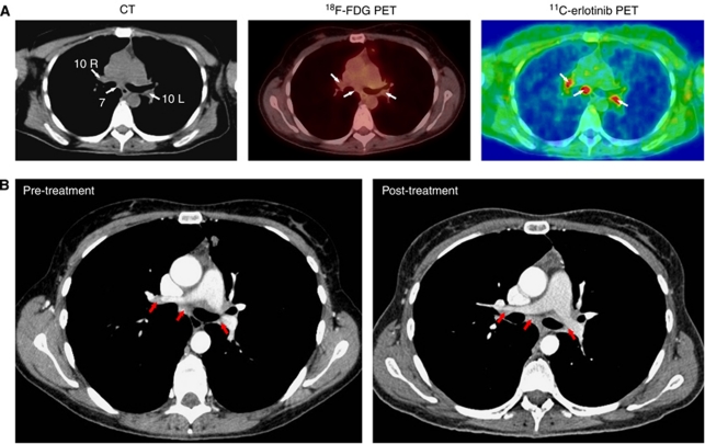
Thirteen patients with NSCLC destined for erlotinib treatment were examined by contrast-enhanced computed tomography (CT), (11)C-erlotinib PET/low-dose CT and (18)F-fluoro-2-deoxy-D-glucose ((18)F-FDG) PET/low-dose CT before start of the erlotinib treatment. After 12 weeks treatment, they were examined by (18)F-FDG PET/contrast-enhanced CT for the assessment of clinical response.
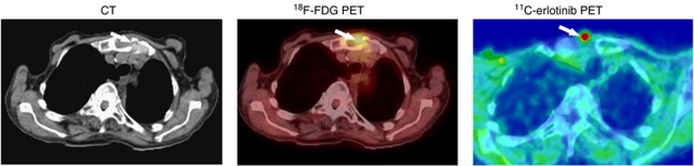
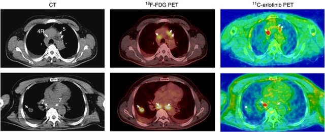
Of the 13 patients included, 4 accumulated (11)C-erlotinib in one or more of their lung tumours or lymph-node metastases. Moreover, (11)C-erlotinib PET/CT identified lesions that were not visible on (18)F-FDG PET/CT. Of the four patients with accumulation of (11)C-erlotinib, one died before follow-up, whereas the other three showed a positive response to erlotinib treatment. Three of the nine patients with no accumulation died before follow-up, four showed progressive disease while two had stable disease after 12 weeks of treatment.
Our data show a potential for (11)C-erlotinib PET/CT for visualizing NSCLC lung tumours, including lymph nodes not identified by (18)F-FDG PET/CT. Large clinical studies are now needed to explore to which extent pre-treatment (11)C-erlotinib PET/CT can predict erlotinib treatment response.
我们之前开发了(11)C-厄洛替尼作为一种新的正电子发射断层扫描(PET)示踪剂,并表明它在小鼠表皮生长因子受体(EGFR)阳性肺癌异种移植瘤中积累。在这里,我们在非小细胞肺癌(NSCLC)患者中进行了一项研究,探讨(11)C-厄洛替尼 PET 作为识别累积厄洛替尼的肺癌肿瘤的潜在方法的可行性。
13 名计划接受厄洛替尼治疗的 NSCLC 患者在开始厄洛替尼治疗前接受了对比增强计算机断层扫描(CT)、(11)C-厄洛替尼 PET/低剂量 CT 和(18)F-氟-2-脱氧-D-葡萄糖((18)F-FDG)PET/低剂量 CT 检查。治疗 12 周后,他们接受(18)F-FDG PET/对比增强 CT 检查以评估临床反应。
在 13 名纳入的患者中,4 名患者的一个或多个肺部肿瘤或淋巴结转移部位有(11)C-厄洛替尼积聚。此外,(11)C-厄洛替尼 PET/CT 还发现了(18)F-FDG PET/CT 无法显示的病变。在 4 名(11)C-厄洛替尼积聚的患者中,1 名患者在随访前死亡,而其余 3 名患者对厄洛替尼治疗有阳性反应。在 9 名无(11)C-厄洛替尼积聚的患者中,有 3 名在随访前死亡,4 名患者疾病进展,2 名患者在治疗 12 周后病情稳定。
我们的数据显示(11)C-厄洛替尼 PET/CT 具有可视化 NSCLC 肺部肿瘤的潜力,包括(18)F-FDG PET/CT 无法识别的淋巴结。现在需要进行大型临床研究,以探讨治疗前(11)C-厄洛替尼 PET/CT 在多大程度上可以预测厄洛替尼治疗反应。