Weintraub Daniel, Doshi Jimit, Koka Deepthi, Davatzikos Christos, Siderowf Andrew D, Duda John E, Wolk David A, Moberg Paul J, Xie Sharon X, Clark Christopher M
Department of Psychiatry, University of Pennsylvania, 3615 Chestnut St, Ste 330, Philadelphia, PA 19104, USA.
Arch Neurol. 2011 Dec;68(12):1562-8. doi: 10.1001/archneurol.2011.725.
To assess regions and patterns of brain atrophy in patients with Parkinson disease (PD) with normal cognition (PD-NC), mild cognitive impairment (PD-MCI), and dementia-level cognitive deficits (PDD).
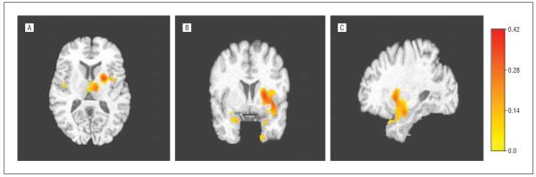
Images were quantified using a region-of-interest approach and voxel-based morphometry analysis. We used a high-dimensional pattern classification approach to delineate brain regions that collectively formed the Spatial Pattern of Abnormalities for Recognition of PDD.
The Parkinson's Disease and Movement Disorders Center at the University of Pennsylvania.
Eighty-four PD patients (61 PD-NC, 12 PD-MCI, and 11 PDD) and 23 healthy control subjects (HCs) underwent magnetic resonance imaging of the brain.
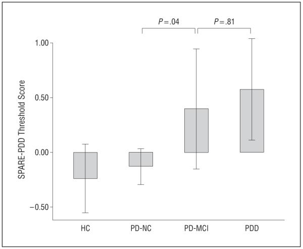
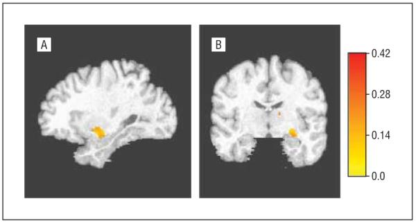
The PD-NC patients did not demonstrate significant brain atrophy compared with HCs. Compared with PD-NC patients, PD-MCI patients had hippocampal atrophy (β = -0.37; P = .001), and PDD patients demonstrated hippocampal (β = -0.32; P = .004) and additional medial temporal lobe atrophy (β = -0.36; P = .003). The PD-MCI patients had a different pattern of atrophy compared with PD-NC patients (P = .04) and a similar pattern to that of PDD patients (P = .81), characterized by hippocampal, prefrontal cortex gray and white matter, occipital lobe gray and white matter, and parietal lobe white matter atrophy. In nondemented PD patients, there was a correlation between memory-encoding performance and hippocampal volume.
Hippocampal atrophy is a biomarker of initial cognitive decline in PD, including impaired memory encoding and storage, suggesting heterogeneity in the neural substrate of memory impairment. Use of a pattern classification approach may allow identification of diffuse regions of cortical gray and white matter atrophy early in the course of cognitive decline.
评估认知正常的帕金森病(PD)患者(PD-NC)、轻度认知障碍(PD-MCI)患者及痴呆水平认知缺陷(PDD)患者的脑萎缩区域和模式。
采用感兴趣区域方法和基于体素的形态计量学分析对图像进行量化。我们使用高维模式分类方法来描绘共同构成PDD识别异常空间模式的脑区。
宾夕法尼亚大学帕金森病与运动障碍中心。
84例PD患者(61例PD-NC、12例PD-MCI和11例PDD)及23名健康对照者(HCs)接受了脑部磁共振成像检查。
与HCs相比,PD-NC患者未表现出明显脑萎缩。与PD-NC患者相比,PD-MCI患者存在海马萎缩(β = -0.37;P = .001),PDD患者表现出海马萎缩(β = -0.32;P = .004)及额外的内侧颞叶萎缩(β = -0.36;P = .003)。与PD-NC患者相比,PD-MCI患者具有不同的萎缩模式(P = .04),与PDD患者的模式相似(P = .81),其特征为海马、前额叶皮质灰质和白质、枕叶灰质和白质以及顶叶白质萎缩。在非痴呆PD患者中,记忆编码表现与海马体积之间存在相关性。
海马萎缩是PD患者初始认知衰退的生物标志物,包括记忆编码和存储受损,提示记忆障碍的神经基质存在异质性。使用模式分类方法可能有助于在认知衰退过程早期识别皮质灰质和白质萎缩的弥散区域。