Brain and Mind Research Program, Central European Institute of Technology, Central European Institute of Technology Masaryk University, Masaryk University, Brno, Czech Republic ; First Department of Neurology, School of Medicine, Masaryk University and St. Anne's University Hospital, Brno, Czech Republic.
Center for Parkinson's disease and Movement Disorder "Fondazione Ospedale San Camillo" - Istituto di Ricovero e Cura a Carattere Scientifico, Venice-Lido, Italy.
PLoS One. 2014 Jan 21;9(1):e85595. doi: 10.1371/journal.pone.0085595. eCollection 2014.
Cortical changes associated with cognitive decline in Parkinson's disease (PD) are not fully explored and require investigations with established diagnostic classification criteria.
We used MRI source-based morphometry to evaluate specific differences in grey matter volume patterns across 4 groups of subjects: healthy controls (HC), PD with normal cognition (PD-NC), PD with mild cognitive impairment (MCI-PD) and PD with dementia (PDD).
We examined 151 consecutive subjects: 25 HC, 75 PD-NC, 29 MCI-PD, and 22 PDD at an Italian and Czech movement disorder centre. Operational diagnostic criteria were applied to classify MCI-PD and PDD. All structural MRI images were processed together in the Czech centre. The spatial independent component analysis was used to assess group differences of local grey matter volume.
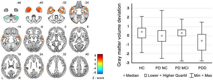
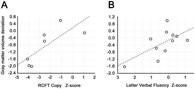
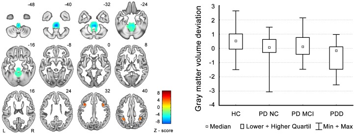
We identified two independent patterns of grey matter volume deviations: a) Reductions in the hippocampus and temporal lobes; b) Decreases in fronto-parietal regions and increases in the midbrain/cerebellum. Both patterns differentiated PDD from all other groups and correlated with visuospatial deficits and letter verbal fluency, respectively. Only the second pattern additionally differentiated PD-NC from HC.
Grey matter changes in PDD involve areas associated with Alzheimer-like pathology while fronto-parietal abnormalities are possibly an early marker of PD cognitive decline. These findings are consistent with a non-linear cognitive progression in PD.
帕金森病(PD)认知能力下降相关的皮质变化尚未得到充分探索,需要使用既定的诊断分类标准进行研究。
我们使用基于 MRI 的形态测量学来评估 4 组受试者的灰质体积模式的特定差异:健康对照(HC)、认知正常的 PD(PD-NC)、轻度认知障碍的 PD(MCI-PD)和痴呆的 PD(PDD)。
我们在意大利和捷克的运动障碍中心检查了 151 名连续受试者:25 名 HC、75 名 PD-NC、29 名 MCI-PD 和 22 名 PDD。操作诊断标准用于分类 MCI-PD 和 PDD。所有结构 MRI 图像都在捷克中心一起进行处理。使用空间独立成分分析来评估局部灰质体积的组间差异。
我们确定了两种灰质体积偏差的独立模式:a)海马体和颞叶减少;b)额顶叶区域减少,中脑/小脑增加。这两种模式均将 PDD 与所有其他组区分开来,分别与视空间缺陷和字母言语流畅性相关。仅第二个模式还将 PD-NC 与 HC 区分开来。
PDD 的灰质变化涉及与阿尔茨海默病样病理学相关的区域,而额顶叶异常可能是 PD 认知能力下降的早期标志物。这些发现与 PD 认知能力的非线性进展一致。