• 文献检索
  • 文档翻译
  • 深度研究
  • 学术资讯
  • Suppr Zotero 插件Zotero 插件
  • 邀请有礼
  • 套餐&价格
  • 历史记录
应用&插件
Suppr Zotero 插件Zotero 插件浏览器插件Mac 客户端Windows 客户端微信小程序
定价
高级版会员购买积分包购买API积分包
服务
文献检索文档翻译深度研究API 文档MCP 服务
关于我们
关于 Suppr公司介绍联系我们用户协议隐私条款
关注我们

Suppr 超能文献

核心技术专利:CN118964589B侵权必究
粤ICP备2023148730 号-1Suppr @ 2026

文献检索

告别复杂PubMed语法,用中文像聊天一样搜索,搜遍4000万医学文献。AI智能推荐,让科研检索更轻松。

立即免费搜索

文件翻译

保留排版,准确专业,支持PDF/Word/PPT等文件格式,支持 12+语言互译。

免费翻译文档

深度研究

AI帮你快速写综述,25分钟生成高质量综述,智能提取关键信息,辅助科研写作。

立即免费体验

Zinn-Haller 环缺血与食蟹猴的青光眼视神经病变。

Ischaemia in the Zinn-Haller circle and glaucomatous optic neuropathy in macaque monkeys.

机构信息

Division of Brain Development and Neuroregeneration, Tokyo Metropolitan Institute of Medical Science, Setagaya-ku, Tokyo, Japan.

出版信息

Br J Ophthalmol. 2012 Apr;96(4):597-603. doi: 10.1136/bjophthalmol-2011-300831. Epub 2012 Jan 4.

DOI:10.1136/bjophthalmol-2011-300831
PMID:22223748
原文链接:https://pmc.ncbi.nlm.nih.gov/articles/PMC3308474/
Abstract

AIMS

To elucidate the morphological features of optic neuropathy in an ischaemic model of glaucoma in macaque monkeys.

METHODS

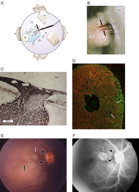
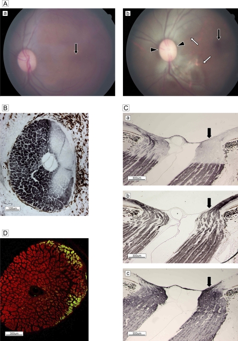
The regional degenerative process was investigated by experimentally occluding the paraoptic branches of the lateral short posterior ciliary artery, that is, the circle of Haller and Zinn, in 11 eyes. Morphological changes in nerve fibres in the lamina cribrosa were evaluated by histopathology, immunocytochemistry and angiography, and the findings were compared with those observed in an aged macaque with spontaneous glaucomatous optic neuropathy.

RESULTS

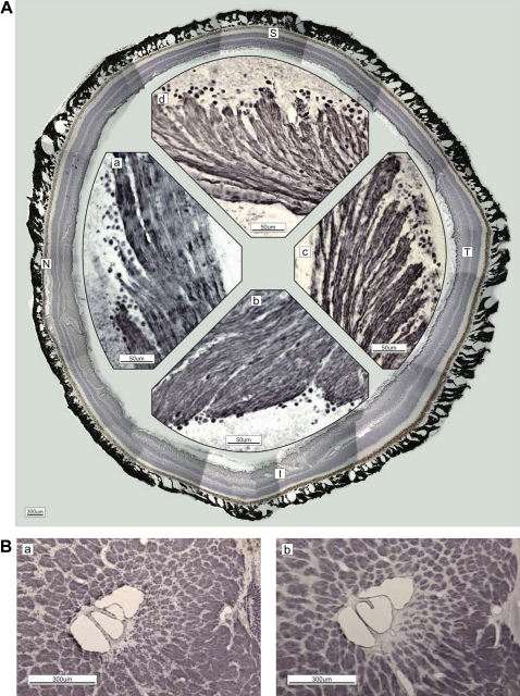
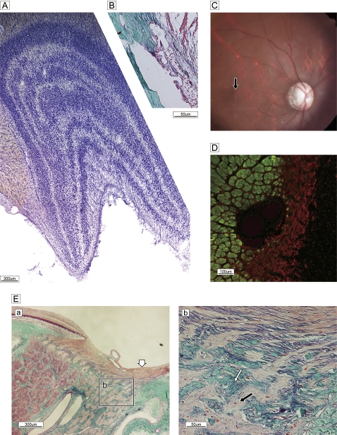
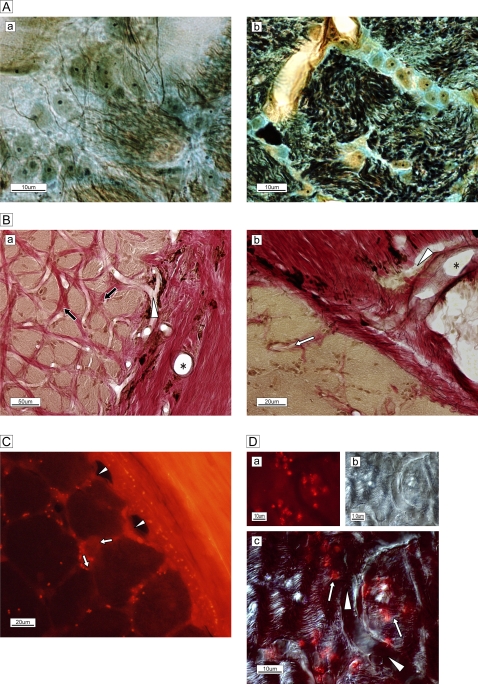
Retinal ganglion cell axons were grouped in bundles and traversed through pores in columns of the lamina cribrosa. The processes of astrocytes extended to the bundles, and capillaries branched in surrounding connective tissue from the circular arterioles. Experimental ischaemia induced time-dependent anoxic deterioration of phosphorylated fibres in the temporal arcuate zone, accompanied by glial proliferation. A monkey with spontaneous visual impairment had nerve fibre loss and gliosis with collagenous proliferation in the temporal hemisphere, suggesting glaucomatous neuropathy.

CONCLUSIONS

Circulatory interference in the circle of Haller and Zinn caused time-dependent deterioration in the area where anoxic segmental degeneration is associated with pathogenesis of open-angle glaucoma.

摘要

目的

阐明猴缺血性青光眼模型中视神经病变的形态学特征。

方法

通过实验性阻断外侧短后睫状动脉的副视支,即 Haller 和 Zinn 环,在 11 只眼中研究区域性退行性过程。通过组织病理学、免疫细胞化学和血管造影评估视盘筛板中神经纤维的形态变化,并将观察结果与自发性青光眼性视神经病变的老年猴进行比较。

结果

视网膜神经节细胞轴突成束穿过筛板的孔,星形胶质细胞的突起延伸到束中,毛细血管从环动脉分支到周围的结缔组织。实验性缺血导致颞弓形区磷酸化纤维的时间依赖性缺氧恶化,伴有胶质增生。一只患有自发性视力障碍的猴子在颞叶半球出现神经纤维丢失和神经胶质增生伴胶原性增殖,提示青光眼性神经病。

结论

Haller 和 Zinn 环的循环干扰导致与开角型青光眼发病机制相关的缺氧节段性变性区域的时间依赖性恶化。

https://cdn.ncbi.nlm.nih.gov/pmc/blobs/4d20/3308474/89102e6305b2/bjophthalmol-2011-300831fig5.jpg
https://cdn.ncbi.nlm.nih.gov/pmc/blobs/4d20/3308474/1698f785d861/bjophthalmol-2011-300831fig1.jpg
https://cdn.ncbi.nlm.nih.gov/pmc/blobs/4d20/3308474/4f8d12baffe6/bjophthalmol-2011-300831fig2.jpg
https://cdn.ncbi.nlm.nih.gov/pmc/blobs/4d20/3308474/d364bae6ec0b/bjophthalmol-2011-300831fig3.jpg
https://cdn.ncbi.nlm.nih.gov/pmc/blobs/4d20/3308474/12e009c17118/bjophthalmol-2011-300831fig4.jpg
https://cdn.ncbi.nlm.nih.gov/pmc/blobs/4d20/3308474/89102e6305b2/bjophthalmol-2011-300831fig5.jpg

相似文献

1
Ischaemia in the Zinn-Haller circle and glaucomatous optic neuropathy in macaque monkeys.Zinn-Haller 环缺血与食蟹猴的青光眼视神经病变。
Br J Ophthalmol. 2012 Apr;96(4):597-603. doi: 10.1136/bjophthalmol-2011-300831. Epub 2012 Jan 4.
2
Thickness of the lamina cribrosa and peripapillary sclera in Rhesus monkeys with nonglaucomatous or glaucomatous optic neuropathy.正常眼压性或青光眼性视神经病变的猕猴的神经节细胞层和视盘周围巩膜厚度。
Acta Ophthalmol. 2011 Aug;89(5):e423-7. doi: 10.1111/j.1755-3768.2011.02121.x. Epub 2011 Feb 18.
3
Detection of Zinn-Haller arterial ring in highly myopic eyes by simultaneous indocyanine green angiography and optical coherence tomography.应用吲哚菁绿血管造影和光相干断层扫描术同时检测高度近视眼中的 Zinn-Haller 动脉环。
Am J Ophthalmol. 2013 May;155(5):920-6. doi: 10.1016/j.ajo.2012.12.010. Epub 2013 Feb 6.
4
Morphologic changes in chronic high-pressure experimental glaucoma in rhesus monkeys.恒河猴慢性高压实验性青光眼的形态学变化
J Glaucoma. 1999 Feb;8(1):56-71.
5
Lamina cribrosa position and Bruch's membrane opening differences between anterior ischemic optic neuropathy and open-angle glaucoma.前部缺血性视神经病变与开角型青光眼之间的筛板位置及布鲁赫膜开口差异
Eur J Ophthalmol. 2019 Mar;29(2):202-209. doi: 10.1177/1120672118782101. Epub 2018 Jun 18.
6
[Lamina cribrosa defect and progress of glaucoma].[筛板缺损与青光眼的进展]
Zhonghua Yan Ke Za Zhi. 2020 Jan 11;56(1):17-20. doi: 10.3760/cma.j.issn.0412-4081.2020.01.007.
7
PERIPAPILLARY ARTERIAL RING OF ZINN-HALLER IN HIGHLY MYOPIC EYES AS DETECTED BY OPTICAL COHERENCE TOMOGRAPHY ANGIOGRAPHY.光学相干断层扫描血管造影术检测高度近视眼中的Zinn-Haller视乳头周围动脉环
Retina. 2017 Feb;37(2):299-304. doi: 10.1097/IAE.0000000000001165.
8
What is glaucomatous optic neuropathy?什么是青光眼性视神经病变?
Jpn J Ophthalmol. 2020 May;64(3):243-249. doi: 10.1007/s10384-020-00736-1. Epub 2020 May 12.
9
Experimental Glaucoma Causes Optic Nerve Head Neural Rim Tissue Compression: A Potentially Important Mechanism of Axon Injury.实验性青光眼导致视神经乳头神经纤维层组织受压:轴突损伤的一个潜在重要机制。
Invest Ophthalmol Vis Sci. 2016 Aug 1;57(10):4403-11. doi: 10.1167/iovs.16-20000.
10
Optic nerve head structure in glaucoma: astrocytes as mediators of axonal damage.青光眼视乳头结构:星形胶质细胞作为轴突损伤的介质
Eye (Lond). 2000 Jun;14 ( Pt 3B):437-44. doi: 10.1038/eye.2000.128.

引用本文的文献

1
Identification of key genes and pathways in scleral extracellular matrix remodeling in glaucoma: Potential therapeutic agents discovered using bioinformatics analysis.利用生物信息学分析鉴定青光眼巩膜细胞外基质重塑中的关键基因和通路:潜在的治疗药物。
Int J Med Sci. 2021 Feb 4;18(7):1554-1565. doi: 10.7150/ijms.52846. eCollection 2021.
2
Progression of glaucomatous optic neuropathy associated with chorioretinal microvascular embolism after intranasal injection of a corticosteroid suspension.鼻内注射皮质类固醇混悬液后与脉络膜视网膜微血管栓塞相关的青光眼性视神经病变进展
Indian J Ophthalmol. 2020 Aug;68(8):1686-1687. doi: 10.4103/ijo.IJO_2332_19.
3

本文引用的文献

1
Pattern of axonal injury in murine myelin oligodendrocyte glycoprotein induced experimental autoimmune encephalomyelitis: implications for multiple sclerosis.小鼠髓鞘少突胶质细胞糖蛋白诱导的实验性自身免疫性脑脊髓炎中的轴突损伤模式:对多发性硬化症的启示
Neurobiol Dis. 2008 May;30(2):162-73. doi: 10.1016/j.nbd.2008.01.001. Epub 2008 Jan 26.
2
Increase in dephosphorylation of the heavy neurofilament subunit in the monkey chronic glaucoma model.猴慢性青光眼模型中重链神经丝亚基去磷酸化增加。
Invest Ophthalmol Vis Sci. 2003 Jan;44(1):154-9. doi: 10.1167/iovs.02-0398.
3
Lack of blood-brain barrier properties in microvessels of the prelaminar optic nerve head.
Focal lamina cribrosa defects and significant peripapillary choroidal thinning in patients with unilateral branch retinal vein occlusion.
单侧视网膜分支静脉阻塞患者的局限性视盘筛板缺陷和显著的视盘周围脉络膜变薄。
PLoS One. 2020 Mar 12;15(3):e0230293. doi: 10.1371/journal.pone.0230293. eCollection 2020.
4
Optic nerve head damage relation to intracranial pressure and corneal properties of eye in glaucoma risk assessment.青光眼风险评估中视神经头损伤与颅内压和眼部角膜特性的关系。
Med Biol Eng Comput. 2019 Jul;57(7):1591-1603. doi: 10.1007/s11517-019-01983-2. Epub 2019 Apr 26.
5
Seeing the Hidden Lamina: Effects of Exsanguination on the Optic Nerve Head.观察潜在的神经层:失血对视神经头的影响。
Invest Ophthalmol Vis Sci. 2018 May 1;59(6):2564-2575. doi: 10.1167/iovs.17-23356.
6
Systemic hypertension is not protective against chronic intraocular pressure elevation in a rodent model.系统性高血压不能预防啮齿动物模型中慢性眼压升高。
Sci Rep. 2018 May 8;8(1):7107. doi: 10.1038/s41598-018-25264-4.
7
In vivo assessment of changes in corneal hysteresis and lamina cribrosa position during acute intraocular pressure elevation in eyes with markedly asymmetrical glaucoma.在明显不对称性青光眼患者的眼中,急性眼压升高期间角膜滞后和筛板位置变化的体内评估。
Clin Ophthalmol. 2018 Mar 15;12:481-492. doi: 10.2147/OPTH.S151532. eCollection 2018.
8
Cerebrospinal Fluid Pressure: Revisiting Factors Influencing Optic Nerve Head Biomechanics.脑脊液压力:重新审视影响视神经头生物力学的因素。
Invest Ophthalmol Vis Sci. 2018 Jan 1;59(1):154-165. doi: 10.1167/iovs.17-22488.
9
Three-Dimensional Strains in Human Posterior Sclera Using Ultrasound Speckle Tracking.使用超声斑点追踪技术测量人眼后巩膜的三维应变
J Biomech Eng. 2016 Feb;138(2):021015. doi: 10.1115/1.4032124.
10
Eye-specific IOP-induced displacements and deformations of human lamina cribrosa.人板层 cribrosa 的特定于眼的 IOP 诱导位移和变形。
Invest Ophthalmol Vis Sci. 2014 Jan 2;55(1):1-15. doi: 10.1167/iovs.13-12724.
视盘前层视神经头微血管缺乏血脑屏障特性。
Invest Ophthalmol Vis Sci. 2001 Apr;42(5):895-901.
4
Zinn-Haller arterial ring observed by ICG angiography in high myopia.通过吲哚菁绿血管造影术在高度近视眼中观察到的辛-哈勒动脉环。
Br J Ophthalmol. 1998 Dec;82(12):1357-62. doi: 10.1136/bjo.82.12.1357.
5
Microvasculature of the anterior optic nerve.视神经前部的微血管系统。
Surv Ophthalmol. 1994 May;38 Suppl:S107-16; discussion S116-7. doi: 10.1016/0039-6257(94)90054-x.
6
Quantitative morphology of human retrolaminar optic nerve vasculature.人视神经管后段血管系统的定量形态学
Invest Ophthalmol Vis Sci. 1994 Oct;35(11):3858-66.
7
Microvasculature of the human optic nerve.人类视神经的微血管系统。
Am J Ophthalmol. 1995 Jul;120(1):92-102. doi: 10.1016/s0002-9394(14)73763-8.
8
Optic nerve damage in human glaucoma. II. The site of injury and susceptibility to damage.人类青光眼的视神经损伤。II. 损伤部位及易损性
Arch Ophthalmol. 1981 Apr;99(4):635-49. doi: 10.1001/archopht.1981.03930010635009.
9
Optic nerve fast axonal transport abnormalities in primates. Occurrence after short posterior ciliary artery occlusion.
Arch Ophthalmol. 1980 Nov;98(11):2018-22. doi: 10.1001/archopht.1980.01020040870017.
10
Blood supply of the optic nerve head and its role in optic atrophy, glaucoma, and oedema of the optic disc.视神经乳头的血液供应及其在视神经萎缩、青光眼和视盘水肿中的作用。
Br J Ophthalmol. 1969 Nov;53(11):721-48. doi: 10.1136/bjo.53.11.721.