TissueVision, Inc., Cambridge, Massachusetts, USA.
Nat Methods. 2012 Jan 15;9(3):255-8. doi: 10.1038/nmeth.1854.
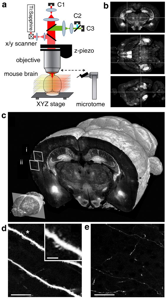
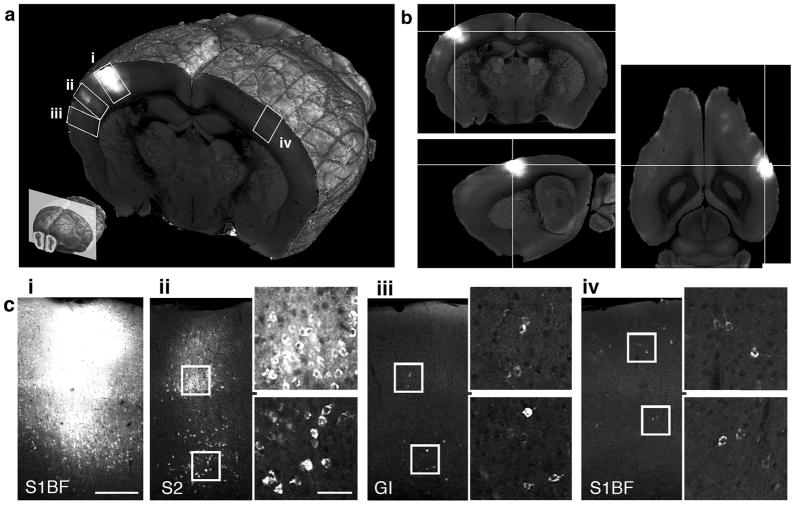
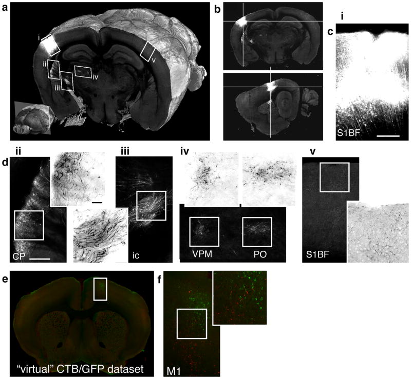
Here we describe an automated method, named serial two-photon (STP) tomography, that achieves high-throughput fluorescence imaging of mouse brains by integrating two-photon microscopy and tissue sectioning. STP tomography generates high-resolution datasets that are free of distortions and can be readily warped in three dimensions, for example, for comparing multiple anatomical tracings. This method opens the door to routine systematic studies of neuroanatomy in mouse models of human brain disorders.
在这里,我们描述了一种自动化方法,称为连续双光子(STP)断层扫描,它通过整合双光子显微镜和组织切片,实现了对小鼠大脑的高通量荧光成像。STP 断层扫描生成的高分辨率数据集没有失真,可以很容易地在三维空间中变形,例如,用于比较多个解剖轨迹。这种方法为在人类大脑疾病的小鼠模型中进行神经解剖学的常规系统研究打开了大门。