• 文献检索
  • 文档翻译
  • 深度研究
  • 学术资讯
  • Suppr Zotero 插件Zotero 插件
  • 邀请有礼
  • 套餐&价格
  • 历史记录
应用&插件
Suppr Zotero 插件Zotero 插件浏览器插件Mac 客户端Windows 客户端微信小程序
定价
高级版会员购买积分包购买API积分包
服务
文献检索文档翻译深度研究API 文档MCP 服务
关于我们
关于 Suppr公司介绍联系我们用户协议隐私条款
关注我们

Suppr 超能文献

核心技术专利:CN118964589B侵权必究
粤ICP备2023148730 号-1Suppr @ 2026

文献检索

告别复杂PubMed语法,用中文像聊天一样搜索,搜遍4000万医学文献。AI智能推荐,让科研检索更轻松。

立即免费搜索

文件翻译

保留排版,准确专业,支持PDF/Word/PPT等文件格式,支持 12+语言互译。

免费翻译文档

深度研究

AI帮你快速写综述,25分钟生成高质量综述,智能提取关键信息,辅助科研写作。

立即免费体验

利用刀扫描显微镜脑图谱进行多尺度小鼠脑微观结构研究。

Multiscale exploration of mouse brain microstructures using the knife-edge scanning microscope brain atlas.

机构信息

Department of Computer Science and Engineering, Texas A&M University College Station, TX, USA.

出版信息

Front Neuroinform. 2011 Nov 22;5:29. doi: 10.3389/fninf.2011.00029. eCollection 2011.

DOI:10.3389/fninf.2011.00029
PMID:22275895
原文链接:https://pmc.ncbi.nlm.nih.gov/articles/PMC3254184/
Abstract

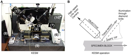
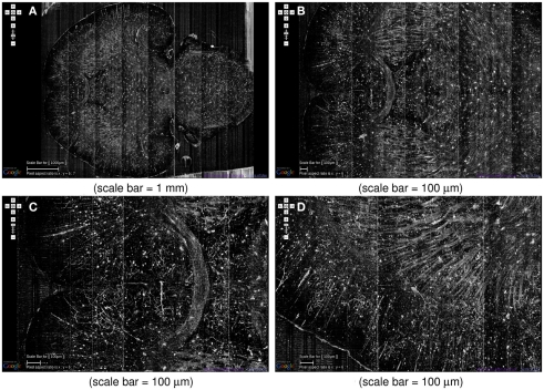
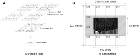
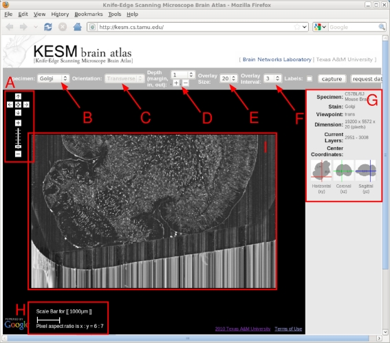
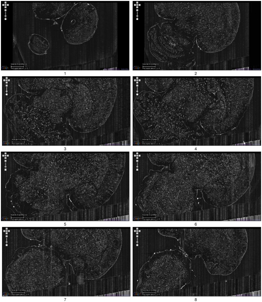
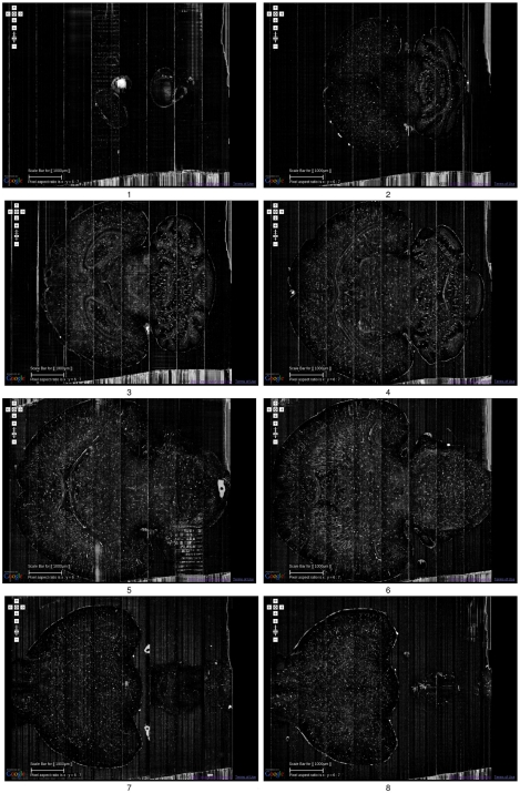
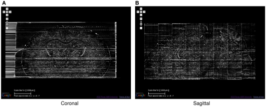
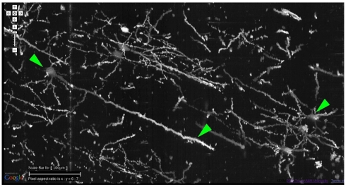
Connectomics is the study of the full connection matrix of the brain. Recent advances in high-throughput, high-resolution 3D microscopy methods have enabled the imaging of whole small animal brains at a sub-micrometer resolution, potentially opening the road to full-blown connectomics research. One of the first such instruments to achieve whole-brain-scale imaging at sub-micrometer resolution is the Knife-Edge Scanning Microscope (KESM). KESM whole-brain data sets now include Golgi (neuronal circuits), Nissl (soma distribution), and India ink (vascular networks). KESM data can contribute greatly to connectomics research, since they fill the gap between lower resolution, large volume imaging methods (such as diffusion MRI) and higher resolution, small volume methods (e.g., serial sectioning electron microscopy). Furthermore, KESM data are by their nature multiscale, ranging from the subcellular to the whole organ scale. Due to this, visualization alone is a huge challenge, before we even start worrying about quantitative connectivity analysis. To solve this issue, we developed a web-based neuroinformatics framework for efficient visualization and analysis of the multiscale KESM data sets. In this paper, we will first provide an overview of KESM, then discuss in detail the KESM data sets and the web-based neuroinformatics framework, which is called the KESM brain atlas (KESMBA). Finally, we will discuss the relevance of the KESMBA to connectomics research, and identify challenges and future directions.

摘要

连接组学是对大脑完整连接矩阵的研究。高通量、高分辨率 3D 显微镜方法的最新进展使得以亚微米分辨率对整个小动物大脑进行成像成为可能,这可能为全面的连接组学研究铺平了道路。第一种实现亚微米分辨率全脑成像的仪器之一是刀刃扫描显微镜(KESM)。KESM 全脑数据集现在包括高尔基(神经元回路)、尼氏(体分布)和印度墨水(血管网络)。KESM 数据可以极大地促进连接组学研究,因为它们填补了较低分辨率、大容量成像方法(如扩散 MRI)和更高分辨率、小体积方法(例如,连续切片电子显微镜)之间的空白。此外,KESM 数据本质上是多尺度的,从亚细胞到整个器官尺度。由于这个原因,仅可视化就是一个巨大的挑战,在我们开始担心定量连接分析之前。为了解决这个问题,我们开发了一个基于网络的神经信息学框架,用于高效可视化和分析多尺度 KESM 数据集。在本文中,我们将首先提供 KESM 的概述,然后详细讨论 KESM 数据集和基于网络的神经信息学框架,称为 KESM 大脑图谱(KESMBA)。最后,我们将讨论 KESMBA 与连接组学研究的相关性,并确定挑战和未来方向。

https://cdn.ncbi.nlm.nih.gov/pmc/blobs/5e13/3254184/1587ce9cc382/fninf-05-00029-g012.jpg
https://cdn.ncbi.nlm.nih.gov/pmc/blobs/5e13/3254184/c4807c802542/fninf-05-00029-g001.jpg
https://cdn.ncbi.nlm.nih.gov/pmc/blobs/5e13/3254184/dbe5c1083a10/fninf-05-00029-g002.jpg
https://cdn.ncbi.nlm.nih.gov/pmc/blobs/5e13/3254184/b07d1b6d1d33/fninf-05-00029-g003.jpg
https://cdn.ncbi.nlm.nih.gov/pmc/blobs/5e13/3254184/63f8c87ec812/fninf-05-00029-g004.jpg
https://cdn.ncbi.nlm.nih.gov/pmc/blobs/5e13/3254184/7e6f63e2db22/fninf-05-00029-g005.jpg
https://cdn.ncbi.nlm.nih.gov/pmc/blobs/5e13/3254184/d1901f7e755b/fninf-05-00029-g006.jpg
https://cdn.ncbi.nlm.nih.gov/pmc/blobs/5e13/3254184/42c520a72663/fninf-05-00029-g007.jpg
https://cdn.ncbi.nlm.nih.gov/pmc/blobs/5e13/3254184/bb8b0af2bf70/fninf-05-00029-g008.jpg
https://cdn.ncbi.nlm.nih.gov/pmc/blobs/5e13/3254184/917f30f1bc72/fninf-05-00029-g009.jpg
https://cdn.ncbi.nlm.nih.gov/pmc/blobs/5e13/3254184/669b5d3c5d65/fninf-05-00029-g010.jpg
https://cdn.ncbi.nlm.nih.gov/pmc/blobs/5e13/3254184/e86f11b4f2ec/fninf-05-00029-g011.jpg
https://cdn.ncbi.nlm.nih.gov/pmc/blobs/5e13/3254184/1587ce9cc382/fninf-05-00029-g012.jpg

相似文献

1
Multiscale exploration of mouse brain microstructures using the knife-edge scanning microscope brain atlas.利用刀扫描显微镜脑图谱进行多尺度小鼠脑微观结构研究。
Front Neuroinform. 2011 Nov 22;5:29. doi: 10.3389/fninf.2011.00029. eCollection 2011.
2
Specimen preparation, imaging, and analysis protocols for knife-edge scanning microscopy.刀口扫描显微镜的样本制备、成像及分析方案
J Vis Exp. 2011 Dec 9(58):3248. doi: 10.3791/3248.
3
Tracing and analysis of the whole mouse brain vasculature with systematic cleaning to remove and consolidate erroneous images.通过系统清理以去除和整合错误图像来追踪和分析整个小鼠脑血管系统。
Annu Int Conf IEEE Eng Med Biol Soc. 2018 Jul;2018:143-146. doi: 10.1109/EMBC.2018.8512215.
4
Fast submicrometer-scale imaging of whole zebrafish using the knife-edge scanning microscope.使用刀口扫描显微镜对整个斑马鱼进行快速亚微米级成像。
Annu Int Conf IEEE Eng Med Biol Soc. 2016 Aug;2016:5901-5904. doi: 10.1109/EMBC.2016.7592071.
5
Mapping the full vascular network in the mouse brain at submicrometer resolution.以亚微米分辨率绘制小鼠大脑中的完整血管网络。
Annu Int Conf IEEE Eng Med Biol Soc. 2017 Jul;2017:3309-3312. doi: 10.1109/EMBC.2017.8037564.
6
Fast macro-scale transmission imaging of microvascular networks using KESM.使用KESM对微血管网络进行快速宏观尺度传输成像。
Biomed Opt Express. 2011 Oct 1;2(10):2888-96. doi: 10.1364/BOE.2.002888. Epub 2011 Sep 29.
7
Robust Cell Detection for Large-Scale 3D Microscopy Using GPU-Accelerated Iterative Voting.使用GPU加速迭代投票进行大规模3D显微镜的稳健细胞检测。
Front Neuroanat. 2018 Apr 26;12:28. doi: 10.3389/fnana.2018.00028. eCollection 2018.
8
Tracing Tubular Structures from Teravoxel-Sized Microscope Images.从万亿体素大小的显微镜图像中追踪管状结构
Annu Int Conf IEEE Eng Med Biol Soc. 2018 Jul;2018:562-565. doi: 10.1109/EMBC.2018.8512288.
9
A Queryable Graph Representation of Vascular Connectivity in the Whole Mouse Brain.
Annu Int Conf IEEE Eng Med Biol Soc. 2019 Jul;2019:256-260. doi: 10.1109/EMBC.2019.8857961.
10
Progress Towards Mammalian Whole-Brain Cellular Connectomics.哺乳动物全脑细胞连接组学的进展
Front Neuroanat. 2016 Jun 30;10:62. doi: 10.3389/fnana.2016.00062. eCollection 2016.

引用本文的文献

1
Merged magnetic resonance and light sheet microscopy of the whole mouse brain.全脑磁共振与光片显微镜融合技术。
Proc Natl Acad Sci U S A. 2023 Apr 25;120(17):e2218617120. doi: 10.1073/pnas.2218617120. Epub 2023 Apr 17.
2
Accurate flow in augmented networks (AFAN): an approach to generating three-dimensional biomimetic microfluidic networks with controlled flow.增强网络中的精确流(AFAN):一种生成具有可控流的三维仿生微流控网络的方法。
Anal Methods. 2019 Jan 7;11(1):8-16. doi: 10.1039/c8ay01798k. Epub 2018 Dec 3.
3
Precise Cerebral Vascular Atlas in Stereotaxic Coordinates of Whole Mouse Brain.

本文引用的文献

1
Fast macro-scale transmission imaging of microvascular networks using KESM.使用KESM对微血管网络进行快速宏观尺度传输成像。
Biomed Opt Express. 2011 Oct 1;2(10):2888-96. doi: 10.1364/BOE.2.002888. Epub 2011 Sep 29.
2
Lateral Connectivity in the Olfactory Bulb is Sparse and Segregated.嗅球的横向连接稀疏且分离。
Front Neural Circuits. 2011 Apr 25;5:5. doi: 10.3389/fncir.2011.00005. eCollection 2011.
3
The DIADEM and beyond.糖尿病防治计划(DIADEM)及其他。 (注:这里DIADEM如果是特定医学领域的专业术语,可能有更准确的固定译法,比如“糖尿病预防与管理数据、教育和监测”之类,具体需结合专业背景确定,这里按字面先给出一种可能译法供参考)
全脑立体定位坐标下的精确脑血管图谱(小鼠脑)
Front Neuroanat. 2017 Dec 19;11:128. doi: 10.3389/fnana.2017.00128. eCollection 2017.
4
Image-guided, Laser-based Fabrication of Vascular-derived Microfluidic Networks.基于图像引导和激光的血管衍生微流控网络制造
J Vis Exp. 2017 Jan 3(119):55101. doi: 10.3791/55101.
5
Possum-A Framework for Three-Dimensional Reconstruction of Brain Images from Serial Sections.Possum:一种用于从连续切片进行脑图像三维重建的框架。
Neuroinformatics. 2016 Jul;14(3):265-78. doi: 10.1007/s12021-015-9286-1.
6
Staining and embedding the whole mouse brain for electron microscopy.对整个小鼠大脑进行染色和包埋用于电子显微镜观察。
Nat Methods. 2012 Dec;9(12):1198-201. doi: 10.1038/nmeth.2213. Epub 2012 Oct 21.
7
Mapping the connectome: multi-level analysis of brain connectivity.绘制连接组:大脑连接性的多层次分析。
Front Neuroinform. 2012 May 1;6:14. doi: 10.3389/fninf.2012.00014. eCollection 2012.
Neuroinformatics. 2011 Sep;9(2-3):99-102. doi: 10.1007/s12021-011-9102-5.
4
Digital atlasing and standardization in the mouse brain.小鼠脑的数字图谱绘制与标准化
PLoS Comput Biol. 2011 Feb 3;7(2):e1001065. doi: 10.1371/journal.pcbi.1001065.
5
Micro-optical sectioning tomography to obtain a high-resolution atlas of the mouse brain.利用微光学切片层析成像技术获取小鼠大脑的高分辨率图谱。
Science. 2010 Dec 3;330(6009):1404-8. doi: 10.1126/science.1191776. Epub 2010 Nov 4.
6
Machines that learn to segment images: a crucial technology for connectomics.能够对图像进行分割的机器:连接组学的关键技术。
Curr Opin Neurobiol. 2010 Oct;20(5):653-66. doi: 10.1016/j.conb.2010.07.004.
7
Waxholm space: an image-based reference for coordinating mouse brain research.瓦克斯霍尔姆空间:用于协调小鼠大脑研究的基于图像的参考标准。
Neuroimage. 2010 Nov 1;53(2):365-72. doi: 10.1016/j.neuroimage.2010.06.067. Epub 2010 Jul 1.
8
NETMORPH: a framework for the stochastic generation of large scale neuronal networks with realistic neuron morphologies.NETMORPH:一个用于随机生成具有逼真神经元形态的大规模神经网络的框架。
Neuroinformatics. 2009 Sep;7(3):195-210. doi: 10.1007/s12021-009-9052-3. Epub 2009 Aug 12.
9
Evaluating automated parameter constraining procedures of neuron models by experimental and surrogate data.通过实验数据和替代数据评估神经元模型的自动参数约束程序。
Biol Cybern. 2008 Nov;99(4-5):371-9. doi: 10.1007/s00422-008-0269-2. Epub 2008 Nov 15.
10
Visualization of cellular and microvascular relationships.细胞与微血管关系的可视化。
IEEE Trans Vis Comput Graph. 2008 Nov-Dec;14(6):1611-8. doi: 10.1109/TVCG.2008.179.