Ophthalmic Research Group, Life and Health Sciences, Aston University, Birmingham B4 7ET, UK.
J Neurol. 2012 Sep;259(9):1832-9. doi: 10.1007/s00415-012-6418-5.
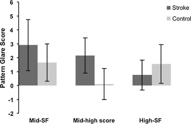
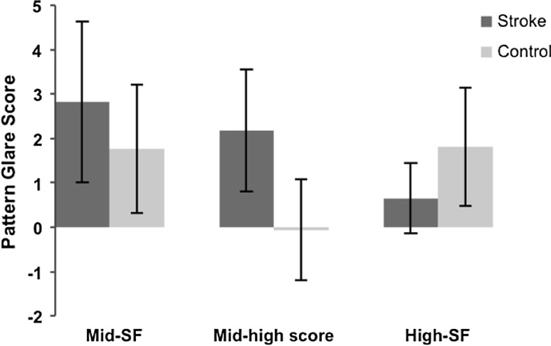
The aim of this work was to measure susceptibility to pattern glare within a stroke group, employing a direct method of assessment. Twenty stroke subjects, aged 38–85 years, were recruited, along with an age-matched control group (n = 20). Assessment of pattern glare susceptibility was undertaken using the pattern glare test. An abnormal degree of pattern glare is present when individuals score[1 on the mid-high spatial frequency difference variable, a relative score that allows for normalization of the subject, or [3 when viewing the mid spatial frequency grating. Stroke subjects demonstrate elevated levels of pattern glare compared to normative data values and a control population, as determined using the pattern glare test. This was most notable when considering the output measure for the mid-high difference variable. The mean score for the mid-high difference variable was 2.15 SD 1.27 for the stroke subjects versus 0.10 SD 1.12 for the control subjects. When considering the mid-high difference variable, 75% of the stroke group recorded an abnormal level of pattern glare compared to 5% in the control group. This study demonstrates an association between stroke subjects and elevated levels of pattern glare. Cortical hyperexcitability has been shown to present following stroke, and this has been proposed as a plausible explanation for the perceptual distortions experienced by individuals susceptible to pattern glare. Further work to assess the benefits of spectral filters in reducing perceptual distortions in stroke patients is currently underway.
本研究旨在采用直接评估方法,测量脑卒中患者对图形眩光的易感性。共招募了 20 名年龄在 38-85 岁的脑卒中患者作为观察组,以及年龄匹配的 20 名健康对照组。采用图形眩光测试评估患者对图形眩光的易感性。如果个体在中高空间频率差异变量上的得分[1,这是一个允许对被试进行归一化的相对分数,或者[3 在观看中空间频率光栅时,则存在异常程度的图形眩光。与正常数据值和对照组相比,脑卒中患者在图形眩光测试中表现出较高水平的图形眩光。当考虑中高差异变量的输出测量值时,这一点最为明显。中高差异变量的平均得分为脑卒中患者组 2.15 SD 1.27,对照组为 0.10 SD 1.12。当考虑中高差异变量时,75%的脑卒中组患者出现图形眩光异常,而对照组为 5%。本研究表明,脑卒中患者与较高水平的图形眩光之间存在关联。脑卒中后皮层兴奋性增加已被证明,这被认为是对图形眩光易感个体所经历的感知扭曲的一种合理解释。目前正在进行进一步的研究,以评估光谱滤光片在减少脑卒中患者感知扭曲方面的益处。