• 文献检索
  • 文档翻译
  • 深度研究
  • 学术资讯
  • Suppr Zotero 插件Zotero 插件
  • 邀请有礼
  • 套餐&价格
  • 历史记录
应用&插件
Suppr Zotero 插件Zotero 插件浏览器插件Mac 客户端Windows 客户端微信小程序
定价
高级版会员购买积分包购买API积分包
服务
文献检索文档翻译深度研究API 文档MCP 服务
关于我们
关于 Suppr公司介绍联系我们用户协议隐私条款
关注我们

Suppr 超能文献

核心技术专利:CN118964589B侵权必究
粤ICP备2023148730 号-1Suppr @ 2026

文献检索

告别复杂PubMed语法,用中文像聊天一样搜索,搜遍4000万医学文献。AI智能推荐,让科研检索更轻松。

立即免费搜索

文件翻译

保留排版,准确专业,支持PDF/Word/PPT等文件格式,支持 12+语言互译。

免费翻译文档

深度研究

AI帮你快速写综述,25分钟生成高质量综述,智能提取关键信息,辅助科研写作。

立即免费体验

ESUR 前列腺磁共振成像指南 2012.

ESUR prostate MR guidelines 2012.

机构信息

Department of Radiology, Radboud University Nijmegen Medical Center, Nijmegen, The Netherlands.

出版信息

Eur Radiol. 2012 Apr;22(4):746-57. doi: 10.1007/s00330-011-2377-y. Epub 2012 Feb 10.

DOI:10.1007/s00330-011-2377-y
PMID:22322308
原文链接:https://pmc.ncbi.nlm.nih.gov/articles/PMC3297750/
Abstract

UNLABELLED

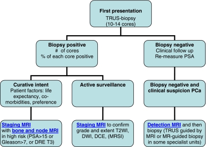
The aim was to develop clinical guidelines for multi-parametric MRI of the prostate by a group of prostate MRI experts from the European Society of Urogenital Radiology (ESUR), based on literature evidence and consensus expert opinion. True evidence-based guidelines could not be formulated, but a compromise, reflected by "minimal" and "optimal" requirements has been made. The scope of these ESUR guidelines is to promulgate high quality MRI in acquisition and evaluation with the correct indications for prostate cancer across the whole of Europe and eventually outside Europe. The guidelines for the optimal technique and three protocols for "detection", "staging" and "node and bone" are presented. The use of endorectal coil vs. pelvic phased array coil and 1.5 vs. 3 T is discussed. Clinical indications and a PI-RADS classification for structured reporting are presented.

KEY POINTS

This report provides guidelines for magnetic resonance imaging (MRI) in prostate cancer. Clinical indications, and minimal and optimal imaging acquisition protocols are provided. A structured reporting system (PI-RADS) is described.

摘要

目的是由欧洲泌尿生殖放射学会(ESUR)的一组前列腺 MRI 专家基于文献证据和共识专家意见,制定前列腺多参数 MRI 的临床指南。由于缺乏真实的循证医学指南,因此制定了一个妥协方案,反映了“最低”和“最佳”要求。这些 ESUR 指南的范围是在整个欧洲甚至欧洲以外地区推广高质量的 MRI 采集和评估,并具有正确的前列腺癌适应证。本文介绍了最佳技术指南以及用于“检测”、“分期”和“淋巴结和骨骼”的三个协议。讨论了直肠内线圈与盆腔相控阵线圈以及 1.5T 与 3T 的使用。还介绍了临床适应证和用于结构化报告的 PI-RADS 分类。

关键点

本报告提供了前列腺癌磁共振成像(MRI)的指南。提供了临床适应证、最低和最佳成像采集方案,并描述了一种结构化报告系统(PI-RADS)。

https://cdn.ncbi.nlm.nih.gov/pmc/blobs/dcfa/3297750/21d68633c4a6/330_2011_2377_Fig3_HTML.jpg
https://cdn.ncbi.nlm.nih.gov/pmc/blobs/dcfa/3297750/9d68dddc05d1/330_2011_2377_Fig1_HTML.jpg
https://cdn.ncbi.nlm.nih.gov/pmc/blobs/dcfa/3297750/66e1dea7dfe8/330_2011_2377_Fig2_HTML.jpg
https://cdn.ncbi.nlm.nih.gov/pmc/blobs/dcfa/3297750/21d68633c4a6/330_2011_2377_Fig3_HTML.jpg
https://cdn.ncbi.nlm.nih.gov/pmc/blobs/dcfa/3297750/9d68dddc05d1/330_2011_2377_Fig1_HTML.jpg
https://cdn.ncbi.nlm.nih.gov/pmc/blobs/dcfa/3297750/66e1dea7dfe8/330_2011_2377_Fig2_HTML.jpg
https://cdn.ncbi.nlm.nih.gov/pmc/blobs/dcfa/3297750/21d68633c4a6/330_2011_2377_Fig3_HTML.jpg

相似文献

1
ESUR prostate MR guidelines 2012.ESUR 前列腺磁共振成像指南 2012.
Eur Radiol. 2012 Apr;22(4):746-57. doi: 10.1007/s00330-011-2377-y. Epub 2012 Feb 10.
2
ESUR prostate MR guidelines.欧洲泌尿生殖放射学会前列腺磁共振成像指南
Eur Radiol. 2013 Aug;23(8):2320-1. doi: 10.1007/s00330-013-2892-0. Epub 2013 May 29.
3
ESUR prostate MR guidelines. Author reply.欧洲泌尿生殖放射学会前列腺磁共振成像指南。作者回复。
Eur Radiol. 2013 Aug;23(8):2322-3. doi: 10.1007/s00330-013-2896-9. Epub 2013 Jun 11.
4
ESUR/ESUI consensus statements on multi-parametric MRI for the detection of clinically significant prostate cancer: quality requirements for image acquisition, interpretation and radiologists' training.ESUR/ESUI 共识声明:多参数 MRI 检测临床显著前列腺癌:图像采集、解读和放射科医生培训的质量要求。
Eur Radiol. 2020 Oct;30(10):5404-5416. doi: 10.1007/s00330-020-06929-z. Epub 2020 May 19.
5
Prostate Cancer: The European Society of Urogenital Radiology Prostate Imaging Reporting and Data System Criteria for Predicting Extraprostatic Extension by Using 3-T Multiparametric MR Imaging.前列腺癌:欧洲泌尿生殖放射学会前列腺成像报告和数据系统标准,用于预测使用 3T 多参数磁共振成像的前列腺外扩展。
Radiology. 2015 Aug;276(2):479-89. doi: 10.1148/radiol.15141412. Epub 2015 Apr 3.
6
European society of urogenital radiology (ESUR) guidelines: MR imaging of pelvic endometriosis.欧洲泌尿生殖放射学会(ESUR)指南:盆腔子宫内膜异位症的磁共振成像
Eur Radiol. 2017 Jul;27(7):2765-2775. doi: 10.1007/s00330-016-4673-z. Epub 2016 Dec 5.
7
Prostate Imaging--An Update.前列腺成像——最新进展
Rofo. 2015 Sep;187(9):751-9. doi: 10.1055/s-0035-1553162. Epub 2015 Jun 26.
8
Use of the Prostate Imaging Reporting and Data System (PI-RADS) for Prostate Cancer Detection with Multiparametric Magnetic Resonance Imaging: A Diagnostic Meta-analysis.使用前列腺影像报告和数据系统(PI-RADS)对前列腺癌进行多参数磁共振成像检测:一项诊断性荟萃分析。
Eur Urol. 2015 Jun;67(6):1112-1121. doi: 10.1016/j.eururo.2014.10.033. Epub 2014 Nov 6.
9
[PI-RADS classification: structured reporting for MRI of the prostate].[前列腺影像报告和数据系统(PI-RADS)分类:前列腺MRI的结构化报告]
Rofo. 2013 Mar;185(3):253-61. doi: 10.1055/s-0032-1330270. Epub 2013 Feb 12.
10
Multiparametric MR imaging of the prostate at 1.5-T without endorectal coil using an 8 channel pelvic phased array: Is it still a viable option?1.5T 无直肠内线圈 8 通道盆腔相控阵线圈前列腺多参数磁共振成像:是否仍然可行?
Radiography (Lond). 2021 May;27(2):459-463. doi: 10.1016/j.radi.2020.10.011. Epub 2020 Nov 2.

引用本文的文献

1
Diagnostic value and external validation of biparametric magnetic resonance imaging radiomics in clinically significant prostate cancer.双参数磁共振成像放射组学在临床显著性前列腺癌中的诊断价值及外部验证
Transl Androl Urol. 2025 Aug 30;14(8):2269-2278. doi: 10.21037/tau-2025-209. Epub 2025 Aug 25.
2
Diagnostic Yield of Fusion-Guided and Randomized Biopsies in Prostate Cancer: Evidence for an Integrated Approach.融合引导活检与随机活检在前列腺癌诊断中的检出率:综合方法的证据
Healthcare (Basel). 2025 Sep 4;13(17):2214. doi: 10.3390/healthcare13172214.
3
Biparametric MRI-based radiomics for differentiating clinically significant prostate cancer among prostate-specific antigen level of gray zone.

本文引用的文献

1
Prospective assessment of prostate cancer aggressiveness using 3-T diffusion-weighted magnetic resonance imaging-guided biopsies versus a systematic 10-core transrectal ultrasound prostate biopsy cohort.前瞻性评估使用 3-T 扩散加权磁共振成像引导活检与系统的 10 核经直肠超声前列腺活检队列对前列腺癌侵袭性的影响。
Eur Urol. 2012 Jan;61(1):177-84. doi: 10.1016/j.eururo.2011.08.042. Epub 2011 Aug 27.
2
Relationship between apparent diffusion coefficients at 3.0-T MR imaging and Gleason grade in peripheral zone prostate cancer.3.0T MRI 下表观扩散系数与前列腺癌外周带 Gleason 分级的关系。
Radiology. 2011 May;259(2):453-61. doi: 10.1148/radiol.11091409.
3
基于双参数MRI的影像组学在前列腺特异性抗原水平处于灰色地带时鉴别临床显著性前列腺癌中的应用
Front Oncol. 2025 Aug 27;15:1615005. doi: 10.3389/fonc.2025.1615005. eCollection 2025.
4
Biparametric vs Multiparametric MRI for Prostate Cancer Diagnosis: The PRIME Diagnostic Clinical Trial.双参数磁共振成像与多参数磁共振成像在前列腺癌诊断中的应用:PRIME诊断临床试验
JAMA. 2025 Sep 10. doi: 10.1001/jama.2025.13722.
5
Prostate MR image segmentation using a multi-stage network approach.使用多阶段网络方法进行前列腺磁共振图像分割。
Int Urol Nephrol. 2025 Sep 5. doi: 10.1007/s11255-025-04763-0.
6
The use of ADC histogram analysis in the diagnosis and determination of aggressiveness of peripheral zone prostate cancer.表观扩散系数(ADC)直方图分析在周围区前列腺癌诊断及侵袭性判定中的应用
Pol J Radiol. 2025 Jul 28;90:e374-e383. doi: 10.5114/pjr/205459. eCollection 2025.
7
Comparison of biparameter and multiparameter MRI in detection of clinically significant prostate cancer across PSA stratifications.双参数和多参数MRI在不同前列腺特异性抗原(PSA)分层中检测临床显著性前列腺癌的比较。
BMC Med Imaging. 2025 Aug 25;25(1):346. doi: 10.1186/s12880-025-01884-x.
8
Improving risk stratification of PI-RADS 3 + 1 lesions of the peripheral zone: expert lexicon of terms, multi-reader performance and contribution of artificial intelligence.改善外周区PI-RADS 3+1病变的风险分层:术语专家词典、多读者表现及人工智能的贡献
Cancer Imaging. 2025 Aug 19;25(1):102. doi: 10.1186/s40644-025-00916-7.
9
Accuracy, intra-, and inter-radiologist variability of PI-RADS v2.1 scoring for clinically significant prostate cancer detection.PI-RADS v2.1评分在检测临床显著性前列腺癌方面的准确性、放射科医生内部及之间的变异性。
Quant Imaging Med Surg. 2025 Aug 1;15(8):7080-7089. doi: 10.21037/qims-2025-37. Epub 2025 Jul 30.
10
Impact of magnetic resonance imaging visibility of prostate cancer on partial gland ablation.前列腺癌磁共振成像可见性对部分腺体消融的影响。
BJUI Compass. 2025 Aug 6;6(8):e70065. doi: 10.1002/bco2.70065. eCollection 2025 Aug.
Prostate cancer: 1HMRS-DCEMR at 3T versus [(18)F]choline PET/CT in the detection of local prostate cancer recurrence in men with biochemical progression after radical retropubic prostatectomy (RRP).
前列腺癌:3T 下 1HMRS-DCEMR 与 [(18)F]胆碱 PET/CT 对根治性经耻骨后前列腺切除术(RRP)后生化复发的男性局部前列腺癌复发的检测比较。
Eur J Radiol. 2012 Apr;81(4):700-8. doi: 10.1016/j.ejrad.2011.01.095. Epub 2011 Feb 17.
4
Areas suspicious for prostate cancer: MR-guided biopsy in patients with at least one transrectal US-guided biopsy with a negative finding--multiparametric MR imaging for detection and biopsy planning.可疑前列腺癌区域:至少进行过一次经直肠超声引导下前列腺活检且结果为阴性的患者——多参数磁共振成像用于检测和活检规划。
Radiology. 2011 Apr;259(1):162-72. doi: 10.1148/radiol.10101251. Epub 2011 Jan 13.
5
Magnetic resonance imaging for the detection, localisation, and characterisation of prostate cancer: recommendations from a European consensus meeting.磁共振成像在前列腺癌的检测、定位和特征描述中的应用:来自欧洲共识会议的建议。
Eur Urol. 2011 Apr;59(4):477-94. doi: 10.1016/j.eururo.2010.12.009. Epub 2010 Dec 21.
6
Clinical utility of apparent diffusion coefficient (ADC) values in patients with prostate cancer: can ADC values contribute to assess the aggressiveness of prostate cancer?表观扩散系数(ADC)值在前列腺癌患者中的临床应用:ADC 值能否有助于评估前列腺癌的侵袭性?
J Magn Reson Imaging. 2011 Jan;33(1):167-72. doi: 10.1002/jmri.22317.
7
Is apparent diffusion coefficient associated with clinical risk scores for prostate cancers that are visible on 3-T MR images?表观扩散系数与在3-T磁共振图像上可见的前列腺癌临床风险评分相关吗?
Radiology. 2011 Feb;258(2):488-95. doi: 10.1148/radiol.10100667. Epub 2010 Dec 21.
8
An exploratory study of endorectal magnetic resonance imaging and spectroscopy of the prostate as preoperative predictive biomarkers of biochemical relapse after radical prostatectomy.经直肠磁共振成像和前列腺波谱分析作为预测前列腺根治性切除术后生化复发的术前预测生物标志物的探索性研究。
J Urol. 2010 Dec;184(6):2320-7. doi: 10.1016/j.juro.2010.07.037. Epub 2010 Oct 16.
9
Prostate cancer: differentiation of central gland cancer from benign prostatic hyperplasia by using diffusion-weighted and dynamic contrast-enhanced MR imaging.前列腺癌:应用扩散加权和动态对比增强磁共振成像鉴别中央腺体癌与前列腺良性增生。
Radiology. 2010 Dec;257(3):715-23. doi: 10.1148/radiol.10100021. Epub 2010 Sep 15.
10
Diffusion-weighted MRI at 3 T for the evaluation of prostate cancer.3.0T 磁共振弥散加权成像在前列腺癌评估中的应用。
AJR Am J Roentgenol. 2010 Jun;194(6):1461-9. doi: 10.2214/AJR.09.3654.