• 文献检索
  • 文档翻译
  • 深度研究
  • 学术资讯
  • Suppr Zotero 插件Zotero 插件
  • 邀请有礼
  • 套餐&价格
  • 历史记录
应用&插件
Suppr Zotero 插件Zotero 插件浏览器插件Mac 客户端Windows 客户端微信小程序
定价
高级版会员购买积分包购买API积分包
服务
文献检索文档翻译深度研究API 文档MCP 服务
关于我们
关于 Suppr公司介绍联系我们用户协议隐私条款
关注我们

Suppr 超能文献

核心技术专利:CN118964589B侵权必究
粤ICP备2023148730 号-1Suppr @ 2026

文献检索

告别复杂PubMed语法,用中文像聊天一样搜索,搜遍4000万医学文献。AI智能推荐,让科研检索更轻松。

立即免费搜索

文件翻译

保留排版,准确专业,支持PDF/Word/PPT等文件格式,支持 12+语言互译。

免费翻译文档

深度研究

AI帮你快速写综述,25分钟生成高质量综述,智能提取关键信息,辅助科研写作。

立即免费体验

足踝部假性肿瘤性软组织病变:影像学综述

Pseudotumoural soft tissue lesions of the foot and ankle: a pictorial review.

作者信息

Van Hul Erik, Vanhoenacker Filip, Van Dyck Pieter, De Schepper Arthur, Parizel Paul M

出版信息

Insights Imaging. 2011 Aug;2(4):439-452. doi: 10.1007/s13244-011-0087-2. Epub 2011 May 1.

DOI:10.1007/s13244-011-0087-2
PMID:22347966
原文链接:https://pmc.ncbi.nlm.nih.gov/articles/PMC3259410/
Abstract

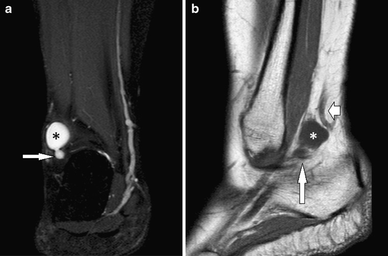
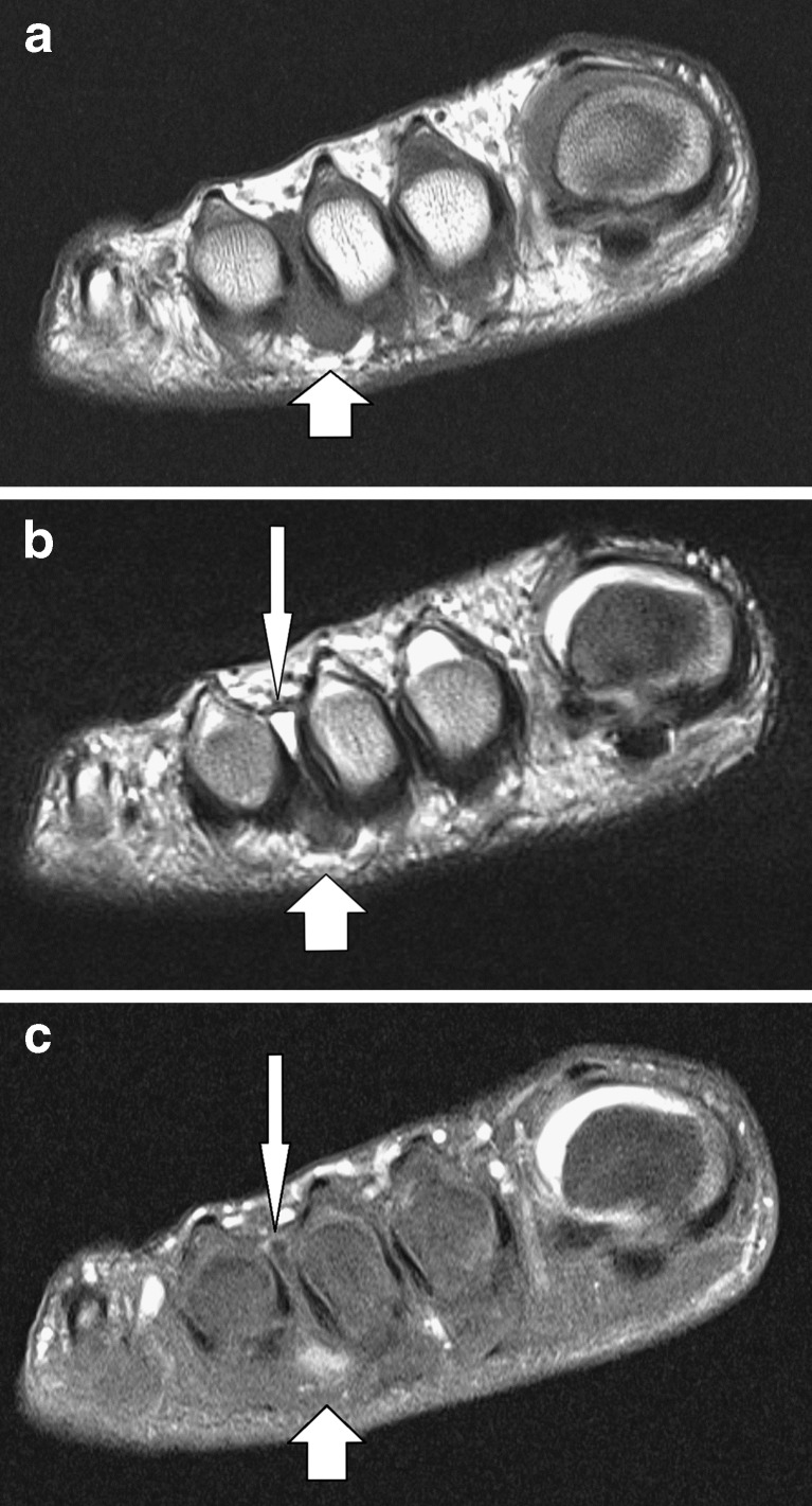
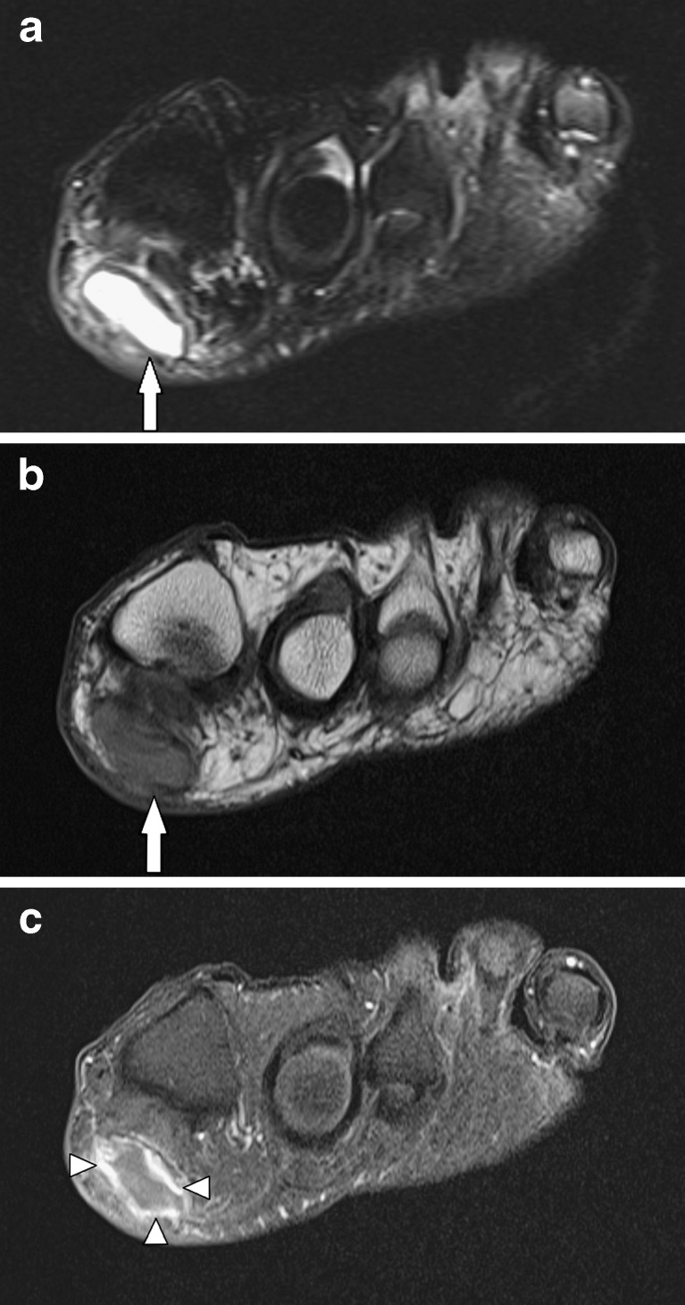
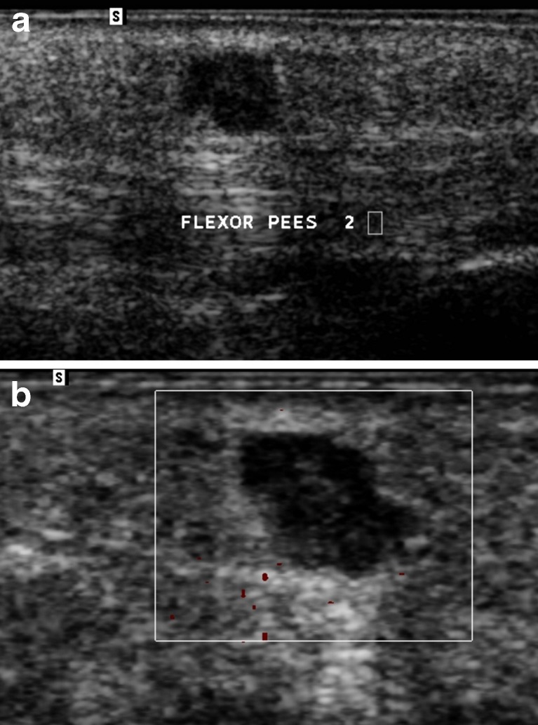
In the foot and ankle region, benign neoplasms and pseudotumoural soft tissue lesions are significantly more frequent than malignant tumours. The pseudotumoural lesions constitute a heterogeneous group, with highly varied aetiology and histopathology. This article reviews the imaging features of the most common pseudotumours of the soft tissues in the foot and ankle. Although the imaging characteristics of several of the lesions discussed are non-specific, combining them with lesion location and clinical features allows the radiologist to suggest a specific diagnosis in most cases.

摘要

在足踝区域,良性肿瘤和假瘤性软组织病变远比恶性肿瘤更为常见。假瘤性病变是一组异质性疾病,其病因和组织病理学差异很大。本文综述了足踝部最常见的软组织假瘤的影像学特征。尽管文中讨论的一些病变的影像学特征不具有特异性,但将它们与病变位置及临床特征相结合,放射科医生在大多数情况下能够做出明确诊断。

https://cdn.ncbi.nlm.nih.gov/pmc/blobs/5fe5/3289016/83b445946414/13244_2011_87_Fig16_HTML.jpg
https://cdn.ncbi.nlm.nih.gov/pmc/blobs/5fe5/3289016/a88d1df22c81/13244_2011_87_Fig1_HTML.jpg
https://cdn.ncbi.nlm.nih.gov/pmc/blobs/5fe5/3289016/a397244df63c/13244_2011_87_Fig2_HTML.jpg
https://cdn.ncbi.nlm.nih.gov/pmc/blobs/5fe5/3289016/a4c83fc1d90e/13244_2011_87_Fig3_HTML.jpg
https://cdn.ncbi.nlm.nih.gov/pmc/blobs/5fe5/3289016/d385018c6d8e/13244_2011_87_Fig4_HTML.jpg
https://cdn.ncbi.nlm.nih.gov/pmc/blobs/5fe5/3289016/098325f9ce4d/13244_2011_87_Fig5_HTML.jpg
https://cdn.ncbi.nlm.nih.gov/pmc/blobs/5fe5/3289016/82107345c519/13244_2011_87_Fig6_HTML.jpg
https://cdn.ncbi.nlm.nih.gov/pmc/blobs/5fe5/3289016/b74bc32b0b55/13244_2011_87_Fig7_HTML.jpg
https://cdn.ncbi.nlm.nih.gov/pmc/blobs/5fe5/3289016/801e354f6e88/13244_2011_87_Fig8_HTML.jpg
https://cdn.ncbi.nlm.nih.gov/pmc/blobs/5fe5/3289016/9212bf87a34c/13244_2011_87_Fig9_HTML.jpg
https://cdn.ncbi.nlm.nih.gov/pmc/blobs/5fe5/3289016/dc84798185aa/13244_2011_87_Fig10_HTML.jpg
https://cdn.ncbi.nlm.nih.gov/pmc/blobs/5fe5/3289016/439bd12097fb/13244_2011_87_Fig11_HTML.jpg
https://cdn.ncbi.nlm.nih.gov/pmc/blobs/5fe5/3289016/de61c865b632/13244_2011_87_Fig12_HTML.jpg
https://cdn.ncbi.nlm.nih.gov/pmc/blobs/5fe5/3289016/32cb05c20908/13244_2011_87_Fig13_HTML.jpg
https://cdn.ncbi.nlm.nih.gov/pmc/blobs/5fe5/3289016/934347e54811/13244_2011_87_Fig14_HTML.jpg
https://cdn.ncbi.nlm.nih.gov/pmc/blobs/5fe5/3289016/4d5b6694dba6/13244_2011_87_Fig15_HTML.jpg
https://cdn.ncbi.nlm.nih.gov/pmc/blobs/5fe5/3289016/83b445946414/13244_2011_87_Fig16_HTML.jpg

相似文献

1
Pseudotumoural soft tissue lesions of the foot and ankle: a pictorial review.足踝部假性肿瘤性软组织病变:影像学综述
Insights Imaging. 2011 Aug;2(4):439-452. doi: 10.1007/s13244-011-0087-2. Epub 2011 May 1.
2
Pseudotumoural soft tissue lesions of the hand and wrist: a pictorial review.手部和腕部的假瘤性软组织病变:影像学综述
Insights Imaging. 2011 Jun;2(3):319-333. doi: 10.1007/s13244-011-0076-5. Epub 2011 Feb 25.
3
Soft tissue masses in the foot and ankle: characteristics on MR Imaging.足踝部软组织肿块:磁共振成像特征
Semin Musculoskelet Radiol. 2005 Sep;9(3):227-42. doi: 10.1055/s-2005-921942.
4
Tumours of the foot and ankle.足踝部肿瘤
Foot (Edinb). 2015 Sep;25(3):164-72. doi: 10.1016/j.foot.2015.06.001. Epub 2015 Jun 10.
5
MR Imaging of Common Soft Tissue Masses in the Foot and Ankle.足踝部常见软组织肿块的磁共振成像
Magn Reson Imaging Clin N Am. 2017 Feb;25(1):159-181. doi: 10.1016/j.mric.2016.08.013.
6
Review of Soft Tissue Masses of the Foot and Ankle: Magnetic Resonance Imaging Features.足踝部软组织肿块的影像学评估:磁共振成像特征
Saudi J Med Med Sci. 2023 Apr-Jun;11(2):117-125. doi: 10.4103/sjmms.sjmms_66_22. Epub 2023 Apr 12.
7
Benign soft tissue tumours of the foot & ankle: A pictorial review.足踝部良性软组织肿瘤:图谱综述
J Clin Orthop Trauma. 2023 Jan 13;37:102105. doi: 10.1016/j.jcot.2023.102105. eCollection 2023 Feb.
8
Malignant tumours of the foot and ankle.足部和踝关节的恶性肿瘤。
Foot Ankle Surg. 2020 Jun;26(4):363-370. doi: 10.1016/j.fas.2019.05.005. Epub 2019 May 11.
9
MRI imaging of soft tissue tumours of the foot and ankle.足踝部软组织肿瘤的磁共振成像
Insights Imaging. 2019 Jun 3;10(1):60. doi: 10.1186/s13244-019-0749-z.
10
[Analysis of clinicopathological characteristics and prognostic factors of foot and ankle soft tissue and bone tumors].足踝部软组织及骨肿瘤的临床病理特征与预后因素分析
Zhonghua Zhong Liu Za Zhi. 2018 Sep 23;40(9):685-689. doi: 10.3760/cma.j.issn.0253-3766.2018.09.010.

引用本文的文献

1
Distribution Patterns of Benign and Malignant Bone and Soft Tissue Tumors and Tumor-like lesions in the Hindfoot and Ankle: A 12.5-year Analysis.后足和踝关节中良性和恶性骨与软组织肿瘤和肿瘤样病变的分布模式:12.5 年分析。
In Vivo. 2024 Sep-Oct;38(5):2383-2389. doi: 10.21873/invivo.13705.
2
Treatments for Morton's neuroma.跖间神经瘤的治疗方法。
Cochrane Database Syst Rev. 2024 Feb 9;2(2):CD014687. doi: 10.1002/14651858.CD014687.pub2.
3
Soft Tissue Sarcomas Mimicking Benign Inflammatory Processes: A Diagnostic Dilemma.酷似良性炎症过程的软组织肉瘤:诊断难题

本文引用的文献

1
Tophaceous gout of the knee: revisiting MRI patterns in 30 patients.膝关节痛风石:30 例患者的 MRI 表现再探讨。
J Clin Rheumatol. 2010 Aug;16(5):209-14. doi: 10.1097/RHU.0b013e3181e92c38.
2
Value of computed tomography arthrography with delayed acquisitions in the work-up of ganglion cysts of the tarsal tunnel: report of three cases.CT 关节造影延迟成像在跗管腱鞘囊肿检查中的价值:三例报告。
Skeletal Radiol. 2010 Apr;39(4):381-6. doi: 10.1007/s00256-009-0864-x. Epub 2010 Jan 29.
3
Pediatric soft-tissue tumors and pseudo-tumors: MR imaging features with pathologic correlation: part 1. Imaging approach, pseudotumors, vascular lesions, and adipocytic tumors.
Mediterr J Rheumatol. 2023 Aug 27;34(4):531-536. doi: 10.31138/mjr.270823.sts. eCollection 2023 Dec.
4
Insights into the Distribution Patterns of Foot and Ankle Tumours: Update on the Perspective of a University Tumour Institute.足踝部肿瘤分布模式的见解:大学肿瘤研究所视角的最新进展
J Clin Med. 2024 Jan 8;13(2):350. doi: 10.3390/jcm13020350.
5
Postoperative clinical and functional outcomes in patients with tumor and tumor-like lesion of foot and ankle.足部和踝部肿瘤和肿瘤样病变患者的术后临床和功能结果。
J Foot Ankle Res. 2022 Oct 14;15(1):75. doi: 10.1186/s13047-022-00582-z.
6
Adventitial bursitis of the dorsal foot presenting as soft tissue masses in patients with autism spectrum disorder.跟蹤研究顯示,腳底筋膜炎可能與痛性痙攣有關。
Skeletal Radiol. 2023 Jun;52(6):1247-1250. doi: 10.1007/s00256-022-04196-1. Epub 2022 Oct 4.
7
Predicting Forefoot-Orthosis Interactions in Rheumatoid Arthritis Using Computational Modelling.使用计算模型预测类风湿性关节炎中前足矫形器的相互作用
Front Bioeng Biotechnol. 2021 Dec 23;9:803725. doi: 10.3389/fbioe.2021.803725. eCollection 2021.
8
Increased frequency of intermetatarsal and submetatarsal bursitis in early rheumatoid arthritis: a large case-controlled MRI study.早期类风湿关节炎中跖间和跖下滑囊炎的频率增加:一项大型病例对照 MRI 研究。
Arthritis Res Ther. 2020 Nov 23;22(1):277. doi: 10.1186/s13075-020-02359-w.
9
Lesions of the heel fat pad.足跟脂肪垫损伤。
Br J Radiol. 2021 Feb 1;94(1118):20200648. doi: 10.1259/bjr.20200648. Epub 2020 Oct 15.
10
Adventitious bursitis in the plantar fat pad of forefoot presenting as a tumoral mass.前足跖脂肪垫的偶然性滑囊炎表现为肿瘤样肿块。
J Radiol Case Rep. 2020 Feb 29;14(2):12-20. doi: 10.3941/jrcr.v14i2.3711. eCollection 2020 Feb.
小儿软组织肿瘤与肿瘤样病变:具有病理对照的磁共振成像特征:第1部分。成像方法、肿瘤样病变、血管性病变及脂肪细胞性肿瘤。
Radiographics. 2009 May-Jun;29(3):887-906. doi: 10.1148/rg.293085168.
4
MR imaging of the diabetic foot.糖尿病足的磁共振成像
Magn Reson Imaging Clin N Am. 2008 Feb;16(1):59-70, vi. doi: 10.1016/j.mric.2008.02.004.
5
Accessory muscles: anatomy, symptoms, and radiologic evaluation.附属肌肉:解剖学、症状及影像学评估
Radiographics. 2008 Mar-Apr;28(2):481-99. doi: 10.1148/rg.282075064.
6
[Soft tissue tumors and pseudotumors of the foot and ankle].[足踝部软组织肿瘤及肿瘤样病变]
J Radiol. 2008 Jan;89(1 Pt 1):21-34. doi: 10.1016/s0221-0363(08)70366-4.
7
Fibrosis and adventitious bursae in plantar fat pad of forefoot: MR imaging findings in asymptomatic volunteers and MR imaging-histologic comparison.前足跖脂肪垫中的纤维化和假性滑囊:无症状志愿者的磁共振成像表现及磁共振成像与组织学对比
Radiology. 2008 Mar;246(3):863-70. doi: 10.1148/radiol.2463070196. Epub 2008 Jan 14.
8
Tumoral calcinosis of the foot with unusual presentation in an 11-year-old boy: a case report and review of literature.11岁男孩足部肿瘤性钙化症的罕见表现:病例报告及文献复习
J Postgrad Med. 2007 Oct-Dec;53(4):247-9. doi: 10.4103/0022-3859.37513.
9
Post-traumatic myositis ossificans circumscripta: an unusually large example.创伤后局限性骨化性肌炎:一个罕见的巨大病例。
J Am Podiatr Med Assoc. 2007 May-Jun;97(3):229-33. doi: 10.7547/0970229.
10
Pseudotumoral appearance of a ruptured epidermal cyst in the foot.足部破裂表皮样囊肿的假瘤样表现。
Skeletal Radiol. 2006 Nov;35(11):867-71. doi: 10.1007/s00256-006-0168-3. Epub 2006 Jun 8.