Interdisciplinary Research Group in Infectious Diseases, Singapore-Massachusetts Institute of Technology Alliance in Research and Technology, Singapore.
PLoS One. 2012;7(2):e31494. doi: 10.1371/journal.pone.0031494. Epub 2012 Feb 15.
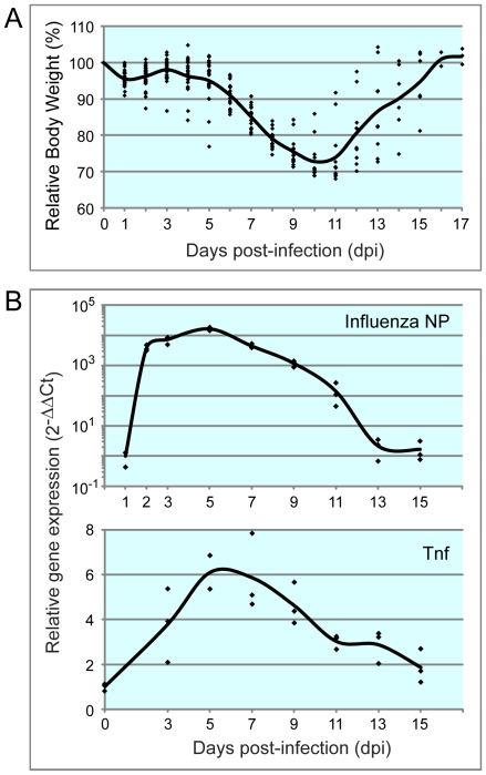
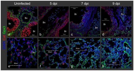
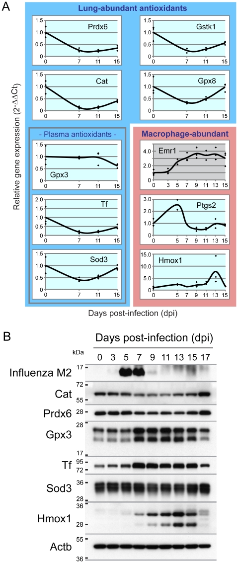
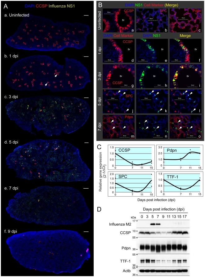
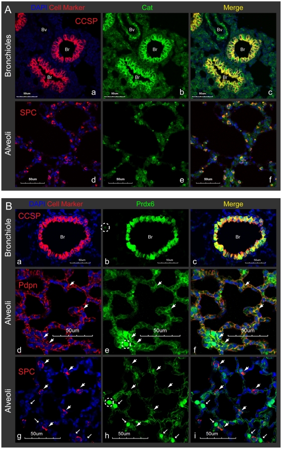
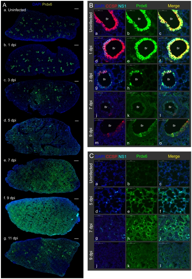
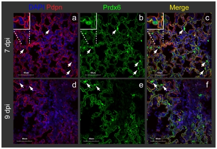
With the incessant challenge of exposure to the air we breathe, lung tissue suffers the highest levels of oxygen tension and thus requires robust antioxidant defenses. Furthermore, following injury or infection, lung tissue faces the additional challenge of inflammation-induced reactive oxygen and nitrogen species (ROS/RNS). Little is known about the identity or distribution of lung antioxidant enzymes under normal conditions or during infection-induced inflammation. Using a mouse model of influenza (H1N1 influenza virus A/PR/8/34 [PR8]) in combination with bioinformatics, we identified seven lung-abundant antioxidant enzymes: Glutathione peroxidase 3 (Gpx3), Superoxide dismutase 3 (Sod3), Transferrin (Tf), peroxyredoxin6 (Prdx6), glutathione S-transferase kappa 1 (Gstk1), Catalase (Cat), and Glutathione peroxidase 8 (Gpx8). Interestingly, despite the demand for antioxidants during inflammation, influenza caused depletion in two key antioxidants: Cat and Prdx6. As Cat is highly expressed in Clara cells, virus-induced Clara cell loss contributes to the depletion in Cat. Prdx6 is also reduced due to Clara cell loss, however there is a coincident increase in Prdx6 levels in the alveoli, resulting in only a subtle reduction of Prdx6 overall. Analogously, Gpx3 shifts from the basement membranes underlying the bronchioles and blood vessels to the alveoli, thus maintaining balanced expression. Taken together, these studies identify key lung antioxidants and reveal their distribution among specific cell types. Furthermore, results show that influenza depletes key antioxidants, and that in some cases there is coincident increased expression, consistent with compensatory expression. Given that oxidative stress is known to be a key risk factor during influenza infection, knowledge about the antioxidant repertoire of lungs, and the spatio-temporal distribution of antioxidants, contributes to our understanding of the underlying mechanisms of influenza-induced morbidity and mortality.
由于不断受到我们所呼吸的空气的暴露,肺部组织承受着最高水平的氧气张力,因此需要强大的抗氧化防御。此外,在受伤或感染后,肺部组织还面临着炎症诱导的活性氧和氮物种(ROS/RNS)的额外挑战。在正常情况下或感染诱导的炎症期间,肺部抗氧化酶的身份或分布知之甚少。我们使用流感(H1N1 流感病毒 A/PR/8/34 [PR8])的小鼠模型与生物信息学相结合,鉴定了七种肺部丰富的抗氧化酶:谷胱甘肽过氧化物酶 3(Gpx3)、超氧化物歧化酶 3(Sod3)、转铁蛋白(Tf)、过氧还蛋白 6(Prdx6)、谷胱甘肽 S-转移酶 kappa 1(Gstk1)、过氧化氢酶(Cat)和谷胱甘肽过氧化物酶 8(Gpx8)。有趣的是,尽管在炎症期间需要抗氧化剂,但流感导致两种关键抗氧化剂的耗竭:Cat 和 Prdx6。由于 Cat 在 Clara 细胞中高度表达,病毒诱导的 Clara 细胞丢失导致 Cat 耗竭。Prdx6 也因 Clara 细胞丢失而减少,但肺泡中 Prdx6 水平同时增加,导致 Prdx6 总体减少不明显。类似地,Gpx3 从细支气管和血管下方的基膜转移到肺泡,从而保持平衡表达。总之,这些研究确定了关键的肺部抗氧化剂,并揭示了它们在特定细胞类型中的分布。此外,结果表明流感会消耗关键的抗氧化剂,在某些情况下,抗氧化剂的表达会同时增加,这与代偿性表达一致。鉴于氧化应激已知是流感感染的一个关键危险因素,了解肺部的抗氧化剂储备以及抗氧化剂的时空分布有助于我们理解流感引起的发病率和死亡率的潜在机制。