Department of Pharmacology, University of Medicine and Dentistry of New Jersey-Robert Wood Johnson Medical School, Piscataway, New Jersey, United States of America.
PLoS One. 2012;7(3):e32542. doi: 10.1371/journal.pone.0032542. Epub 2012 Mar 1.
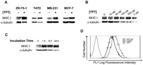
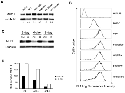
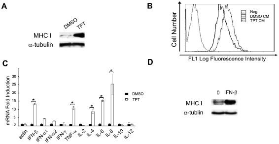
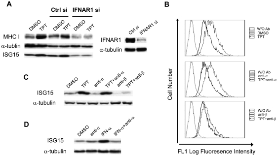
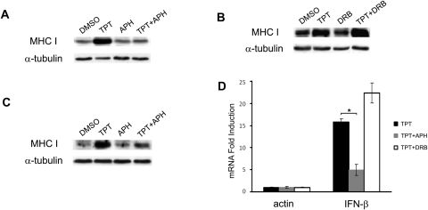
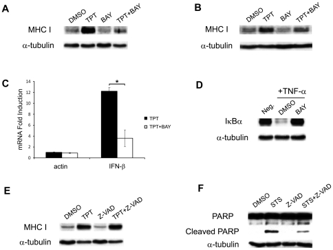
Low doses of anticancer drugs have been shown to enhance antitumor immune response and increase the efficacy of immunotherapy. The molecular basis for such effects remains elusive, although selective depletion of T regulatory cells has been demonstrated. In the current studies, we demonstrate that topotecan (TPT), a topoisomerase I-targeting drug with a well-defined mechanism of action, stimulates major histocompatibility complex class I (MHC I) expression in breast cancer cells through elevated expression/secretion of interferon-β (IFN-β) and activation of type I IFN signaling. First, we show that TPT treatment elevates the expression of both total and cell-surface MHC I in breast cancer cells. Second, conditioned media from TPT-treated breast cancer ZR-75-1 cells induce elevated expression of cell-surface MHC I in drug-naïve recipient cells, suggesting the involvement of cytokines and/or other secreted molecules. Consistently, TPT-treated cells exhibit elevated expression of multiple cytokines such as IFN-β, TNF-α, IL-6 and IL-8. Third, either knocking down the type I interferon receptor subunit 1 (IFNAR1) or addition of neutralizing antibody against IFN-β results in reduced MHC I expression in TPT-treated cells. Together, these results suggest that TPT induces increased IFN-β autocrine/paracrine signaling through type I IFN receptor, resulting in the elevated MHC I expression in tumor cells. Studies have also demonstrated that other chemotherapeutic agents (e.g. etoposide, cisplatin, paclitaxel and vinblastine) similarly induce increased IFN-β secretion and elevated MHC I expression. In addition, conditioned media from γ-irradiated donor cells are shown to induce IFN-β-dependent MHC I expression in unirradiated recipient cells. In the aggregate, our results suggest that many cancer therapeutics induce elevated tumor antigen presentation through MHC I, which could represent a common mechanism for enhanced antitumor immune response through T cell cytotoxicity during metronomic chemotherapy, as well as increased efficacy of combined chemo- (or radio-)/immuno-therapy.
低剂量的抗癌药物已被证明可以增强抗肿瘤免疫反应并提高免疫疗法的疗效。尽管已经证明选择性耗尽 T 调节细胞具有这种作用,但这种作用的分子基础仍然难以捉摸。在当前的研究中,我们证明拓扑替康(TPT),一种作用机制明确的拓扑异构酶 I 靶向药物,通过上调干扰素-β(IFN-β)的表达/分泌并激活 I 型 IFN 信号转导,刺激乳腺癌细胞主要组织相容性复合体 I(MHC I)的表达。首先,我们表明 TPT 处理会提高乳腺癌细胞中总 MHC I 和细胞表面 MHC I 的表达。其次,来自 TPT 处理的乳腺癌 ZR-75-1 细胞的条件培养基在药物-naive 受体细胞中诱导细胞表面 MHC I 的表达升高,表明细胞因子和/或其他分泌分子的参与。一致地,TPT 处理的细胞表现出多种细胞因子如 IFN-β、TNF-α、IL-6 和 IL-8 的表达升高。第三,敲低 I 型干扰素受体亚基 1(IFNAR1)或添加针对 IFN-β 的中和抗体可导致 TPT 处理的细胞中 MHC I 表达减少。总之,这些结果表明 TPT 通过 I 型 IFN 受体诱导增加的 IFN-β 自分泌/旁分泌信号,导致肿瘤细胞中 MHC I 的表达升高。研究还表明,其他化疗药物(如依托泊苷、顺铂、紫杉醇和长春碱)同样诱导增加的 IFN-β 分泌和升高的 MHC I 表达。此外,来自γ-辐照供体细胞的条件培养基被证明可在未辐照的受体细胞中诱导 IFN-β 依赖性 MHC I 表达。总之,我们的结果表明,许多癌症疗法通过 MHC I 诱导升高的肿瘤抗原呈递,这可能代表通过低剂量化疗期间 T 细胞细胞毒性增强抗肿瘤免疫反应以及联合化疗(或放射)/免疫疗法增效的共同机制。