Institute of Physical Chemistry and Abbe Center of Photonics, University of Jena, Helmholtzweg 4, 07743, Jena, Germany.
Anal Bioanal Chem. 2012 May;403(3):745-53. doi: 10.1007/s00216-012-5887-9. Epub 2012 Mar 8.
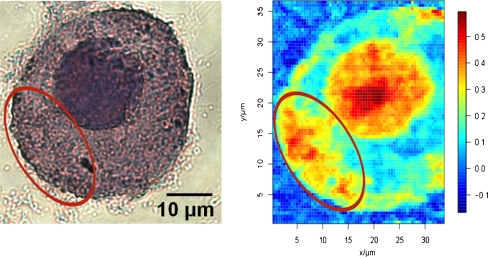
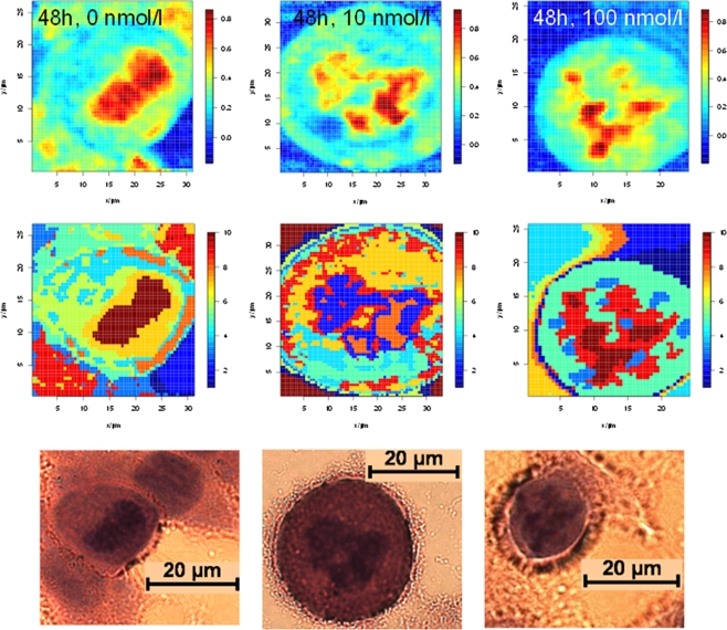
Chemotherapies feature a low success rate of about 25%, and therefore, the choice of the most effective cytostatic drug for the individual patient and monitoring the efficiency of an ongoing chemotherapy are important steps towards personalized therapy. Thereby, an objective method able to differentiate between treated and untreated cancer cells would be essential. In this study, we provide molecular insights into Docetaxel-induced effects in MCF-7 cells, as a model system for adenocarcinoma, by means of Raman microspectroscopy combined with powerful chemometric methods. The analysis of the Raman data is divided into two steps. In the first part, the morphology of cell organelles, e.g. the cell nucleus has been visualized by analysing the Raman spectra with k-means cluster analysis and artificial neural networks and compared to the histopathologic gold standard method hematoxylin and eosin staining. This comparison showed that Raman microscopy is capable of displaying the cell morphology; however, this is in contrast to hematoxylin and eosin staining label free and can therefore be applied potentially in vivo. Because Docetaxel is a drug acting within the cell nucleus, Raman spectra originating from the cell nucleus region were further investigated in a next step. Thereby we were able to differentiate treated from untreated MCF-7 cells and to quantify the cell-drug response by utilizing linear discriminant analysis models.
化疗的成功率约为 25%,因此,选择最有效的细胞抑制剂药物对个体患者进行治疗,并监测正在进行的化疗的效率,是实现个体化治疗的重要步骤。因此,需要有一种能够区分治疗和未治疗癌细胞的客观方法。在这项研究中,我们通过拉曼显微镜与强大的化学计量学方法相结合,提供了多西紫杉醇诱导 MCF-7 细胞(腺癌模型系统)效应的分子见解。拉曼数据分析分为两个步骤。在第一部分中,通过使用 k-均值聚类分析和人工神经网络分析对细胞细胞器的形态(例如细胞核)进行了分析,并与组织病理学金标准方法——苏木精和伊红染色进行了比较。该比较表明,拉曼显微镜能够显示细胞形态;然而,与苏木精和伊红染色不同的是,它是无标记的,因此可以潜在地应用于体内。由于多西紫杉醇是一种在细胞核内起作用的药物,因此在下一步中进一步研究了来自细胞核区域的拉曼光谱。由此,我们能够区分处理过的和未处理的 MCF-7 细胞,并通过利用线性判别分析模型来量化细胞-药物反应。