Coulter Department of Biomedical Engineering Georgia Tech and Emory University, Atlanta, GA 30322, USA.
Neuroimage. 2012 Aug 15;62(2):1103-8. doi: 10.1016/j.neuroimage.2012.03.005. Epub 2012 Mar 9.
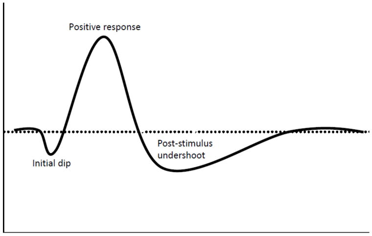
Over the past 20 years much attention has been given to characterizing the spatial accuracy of fMRI based signals and to techniques that improve on its co-localization with neuronal activity. While the vast majority of fMRI studies have always used the conventional positive BOLD signal, alternative contrast options have demonstrated superior spatial specificity. One of these options surfaced shortly after the initial BOLD fMRI demonstrations and was motivated by optical imaging studies which revealed an early signal change that was much smaller but spatially more specific than the delayed positive response. This early signal change was attributed to oxygenation changes prior to any subsequent blood flow increases. After observation of this biphasic hemodynamic response in fMRI, because this early response resulted in a small MR signal decrease prior to the onset of the large signal increase, it became known as the "initial dip". While the initial dip in fMRI was subsequently reported by many studies, including those in humans, monkeys, and cats, there were conflicting views about the associated mechanisms and whether it could be generalized across brain regions or species, in addition to whether or not it would prove fruitful for neuroscience. These discrepancies, along with the implications that the initial dip might increase the spatial specificity of BOLD fMRI from 2 to 3mm to something more closely associated with neural activity, resulted in lot of buzz and controversy in the community for many years. In this review, the authors provide an account of the story of the initial dip in MR based functional imaging from the Minnesota perspective, where the first demonstrations, characterizations, and applications of the initial dip commenced.
在过去的 20 年中,人们对 fMRI 基于信号的空间精度及其提高与神经元活动的局部化的技术给予了极大的关注。尽管绝大多数 fMRI 研究一直使用传统的正 BOLD 信号,但替代对比方案已证明具有更高的空间特异性。其中一种选择方案在最初的 BOLD fMRI 演示之后不久就出现了,其灵感来自于光学成像研究,该研究揭示了一种早期信号变化,其幅度较小,但比延迟的正响应具有更高的空间特异性。这种早期信号变化归因于任何后续血流增加之前的氧合变化。在 fMRI 中观察到这种双相血液动力学反应之后,由于这种早期反应在大信号增加之前导致了较小的 MR 信号降低,因此被称为“初始下降”。尽管许多研究(包括人类,猴子和猫的研究)随后都报道了 fMRI 中的初始下降,但关于相关机制以及它是否可以在大脑区域或物种之间推广,以及它是否对神经科学是否有用,存在相互矛盾的观点。这些差异以及初始下降可能会使 BOLD fMRI 的空间特异性从 2 毫米提高到 3 毫米,或者更紧密地与神经活动相关联的含义,在过去的许多年中在该领域引起了很多关注和争议。在这篇综述中,作者从明尼苏达州的角度讲述了基于磁共振的功能成像中初始下降的故事,最初的演示,特征描述和初始下降的应用都始于明尼苏达州。