• 文献检索
  • 文档翻译
  • 深度研究
  • 学术资讯
  • Suppr Zotero 插件Zotero 插件
  • 邀请有礼
  • 套餐&价格
  • 历史记录
应用&插件
Suppr Zotero 插件Zotero 插件浏览器插件Mac 客户端Windows 客户端微信小程序
定价
高级版会员购买积分包购买API积分包
服务
文献检索文档翻译深度研究API 文档MCP 服务
关于我们
关于 Suppr公司介绍联系我们用户协议隐私条款
关注我们

Suppr 超能文献

核心技术专利:CN118964589B侵权必究
粤ICP备2023148730 号-1Suppr @ 2026

文献检索

告别复杂PubMed语法,用中文像聊天一样搜索,搜遍4000万医学文献。AI智能推荐,让科研检索更轻松。

立即免费搜索

文件翻译

保留排版,准确专业,支持PDF/Word/PPT等文件格式,支持 12+语言互译。

免费翻译文档

深度研究

AI帮你快速写综述,25分钟生成高质量综述,智能提取关键信息,辅助科研写作。

立即免费体验

数字断层合成术在周围神经鞘瘤评估中的应用:与普通放射摄影相比的患者照射量和图像质量比较。

Digital tomosynthesis for PNS evaluation: comparisons of patient exposure and image quality with plain radiography.

机构信息

Department of Radiology, Bundang Jesaeng General Hospital, Sungnam, Korea.

出版信息

Korean J Radiol. 2012 Mar-Apr;13(2):136-43. doi: 10.3348/kjr.2012.13.2.136. Epub 2012 Mar 7.

DOI:10.3348/kjr.2012.13.2.136
PMID:22438680
原文链接:https://pmc.ncbi.nlm.nih.gov/articles/PMC3303896/
Abstract

OBJECTIVE

We investigated low dose digital tomosynthesis (DT) for the evaluation of the paranasal sinus (PNS), and compared its diagnostic accuracy with a PNS radiography series (XR).

MATERIALS AND METHODS

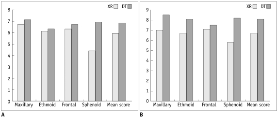
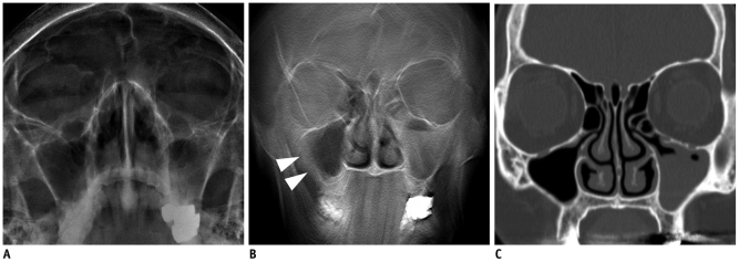
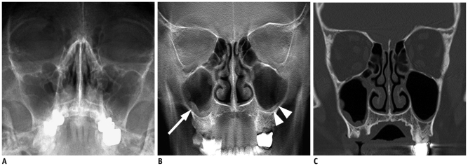
We enrolled 43 patients for whom XR, PNS DT, and OMU CT were performed. We measured effective doses (EDs) of XR, DT, and OMU CT using Monte Carlo simulation software. Two radiologists performed independent observation of both XR and DT. For seven PNSs, they scored anatomic conspicuity of sinuses and confidence on the presence of sinusitis using nine point scales. OMU CT was observed by the third radiologist and the findings were regarded as reference standard. We compared scores for conspicuity and sinusitis confidence between XR and DT.

RESULTS

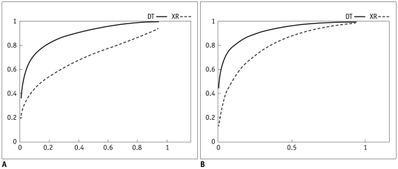
Mean EDs were 29 ± 6 µSv, 48 ± 10 µSv, and 980 ± 250 µSv, respectively, for XR, DT, and CT. Mean scores for conspicuity were 6.3 and 7.4, respectively, for XR and DT. Sensitivity per patient basis for sinusitis detection were 52% and 96%, respectively, for XR and DT in observer 1 (p = 0.001) and 80% and 92% for observer 2 (p = 0.25). Specificities for sinusitis exclusion were 100% for both XR and DT for observer 1 and 89% and 100% for observer 2 (p = 0.50). Accuracies for sinusitis diagnosis were 72% and 98%, respectively, for XR and DT for observer 1 (p = 0.001) and 84% and 95% for observer 2 (p = 0.125).

CONCLUSION

Patient radiation dose from low dose DT is comparable with that of PNS XR. Diagnostic sensitivity of DT for sinusitis was superior to PNS XR.

摘要

目的

我们研究了低剂量数字断层融合摄影术(DT)在鼻窦(PNS)评估中的应用,并将其与 PNS 射线照相系列(XR)的诊断准确性进行了比较。

材料与方法

我们纳入了 43 名患者,他们接受了 XR、PNS DT 和 OMU CT 检查。我们使用蒙特卡罗模拟软件测量了 XR、DT 和 OMU CT 的有效剂量(ED)。两位放射科医生分别对 XR 和 DT 进行独立观察。对于七个鼻窦,他们使用九点量表对鼻窦的解剖显影程度和存在鼻窦炎的信心进行评分。第三位放射科医生观察 OMU CT,并将结果视为参考标准。我们比较了 XR 和 DT 对显影程度和鼻窦炎信心的评分。

结果

XR、DT 和 CT 的平均 ED 分别为 29 ± 6 µSv、48 ± 10 µSv 和 980 ± 250 µSv。XR 和 DT 的平均显影程度评分分别为 6.3 和 7.4。观察者 1 对鼻窦炎检测的敏感度分别为 XR 的 52%和 DT 的 96%(p = 0.001),观察者 2 分别为 80%和 92%(p = 0.25)。对于鼻窦炎的排除,XR 和 DT 的特异度均为 100%,观察者 1 为 89%和 100%,观察者 2 为 89%和 100%(p = 0.50)。对于鼻窦炎的诊断准确性,观察者 1 为 XR 的 72%和 DT 的 98%(p = 0.001),观察者 2 为 84%和 DT 的 95%(p = 0.125)。

结论

低剂量 DT 的患者辐射剂量与 PNS XR 相当。DT 对鼻窦炎的诊断敏感性优于 PNS XR。

https://cdn.ncbi.nlm.nih.gov/pmc/blobs/f6d3/3303896/037ce5d7b948/kjr-13-136-g004.jpg
https://cdn.ncbi.nlm.nih.gov/pmc/blobs/f6d3/3303896/4f50c2983a65/kjr-13-136-g001.jpg
https://cdn.ncbi.nlm.nih.gov/pmc/blobs/f6d3/3303896/ad44cbbb433e/kjr-13-136-g002.jpg
https://cdn.ncbi.nlm.nih.gov/pmc/blobs/f6d3/3303896/bf2d92ce1711/kjr-13-136-g003.jpg
https://cdn.ncbi.nlm.nih.gov/pmc/blobs/f6d3/3303896/037ce5d7b948/kjr-13-136-g004.jpg

相似文献

1
Digital tomosynthesis for PNS evaluation: comparisons of patient exposure and image quality with plain radiography.数字断层合成术在周围神经鞘瘤评估中的应用:与普通放射摄影相比的患者照射量和图像质量比较。
Korean J Radiol. 2012 Mar-Apr;13(2):136-43. doi: 10.3348/kjr.2012.13.2.136. Epub 2012 Mar 7.
2
Detection of paranasal sinus opacification with digital tomosynthesis radiography: a clinical pilot study.数字断层合成摄影术检测鼻窦混浊:一项临床初步研究。
J Comput Assist Tomogr. 2013 Mar-Apr;37(2):252-6. doi: 10.1097/RCT.0b013e31827c0cb1.
3
[Sinus radiography and low-dose CT in the diagnosis of acute sinusitis. ].[鼻窦造影和低剂量CT在急性鼻窦炎诊断中的应用]
Tidsskr Nor Laegeforen. 2003 Dec 4;123(23):3362-4.
4
Radiation Dose Reduction in Paranasal Sinus CT: With Feasibility of Iterative Reconstruction Technique.鼻旁窦CT辐射剂量降低:迭代重建技术的可行性研究
Otolaryngol Head Neck Surg. 2016 Dec;155(6):982-987. doi: 10.1177/0194599816664335. Epub 2016 Aug 23.
5
Reducing the radiation dose for low-dose CT of the paranasal sinuses using iterative reconstruction: feasibility and image quality.使用迭代重建降低鼻窦低剂量 CT 的辐射剂量:可行性和图像质量。
Eur J Radiol. 2012 Sep;81(9):2246-50. doi: 10.1016/j.ejrad.2011.05.002. Epub 2011 Jun 12.
6
Comparison of sinus x-rays with computed tomography scans in acute sinusitis.急性鼻窦炎中鼻窦X光片与计算机断层扫描的比较。
Acad Emerg Med. 1994 May-Jun;1(3):235-9. doi: 10.1111/j.1553-2712.1994.tb02437.x.
7
Digital image analysis of radio-opacities in the paranasal sinuses using computed radiography.
Eur Arch Otorhinolaryngol. 1991;248(6):326-30. doi: 10.1007/BF00169022.
8
Conventional sinus radiography compared with CT in the diagnosis of acute sinusitis.传统鼻窦X线摄影与CT在急性鼻窦炎诊断中的比较。
Dentomaxillofac Radiol. 2003 Jan;32(1):60-2. doi: 10.1259/dmfr/65139094.
9
Digital tomosynthesis of the chest: utility for detection of lung metastasis in patients with colorectal cancer.胸部数字断层合成术:用于检测结直肠癌患者肺部转移的效用。
Clin Radiol. 2012 Mar;67(3):232-8. doi: 10.1016/j.crad.2011.08.017. Epub 2011 Sep 21.
10
Radiation dose of digital tomosynthesis for sinonasal examination: comparison with multi-detector CT.鼻窦检查数字断层融合摄影的辐射剂量:与多探测器 CT 的比较。
Eur J Radiol. 2012 Jun;81(6):1140-5. doi: 10.1016/j.ejrad.2011.03.064. Epub 2011 Apr 22.

引用本文的文献

1
Diagnostic yield of dental radiography and digital tomosynthesis for the identification of anatomic structures in dogs.牙科X线摄影和数字断层合成技术在识别犬类解剖结构中的诊断效能。
Front Vet Sci. 2024 Nov 20;11:1489239. doi: 10.3389/fvets.2024.1489239. eCollection 2024.
2
Digital Tomosynthesis versus Conventional Radiography for Evaluating Osteonecrosis of the Femoral Head.数字断层融合摄影术与常规放射摄影术评估股骨头坏死的比较。
Korean J Radiol. 2021 Dec;22(12):2026-2033. doi: 10.3348/kjr.2021.0058. Epub 2021 Sep 13.
3
Selection and Reporting of Statistical Methods to Assess Reliability of a Diagnostic Test: Conformity to Recommended Methods in a Peer-Reviewed Journal.

本文引用的文献

1
Pulmonary mycobacterial disease: diagnostic performance of low-dose digital tomosynthesis as compared with chest radiography.肺部分枝杆菌病:与胸部 X 线摄影相比,低剂量数字断层合成术的诊断性能。
Radiology. 2010 Oct;257(1):269-77. doi: 10.1148/radiol.10100303.
2
X-ray tomosynthesis: a review of its use for breast and chest imaging.X线断层合成成像:用于乳腺和胸部成像的综述
Radiat Prot Dosimetry. 2010 Apr-May;139(1-3):100-7. doi: 10.1093/rpd/ncq099. Epub 2010 Mar 16.
3
Overview of two years of clinical experience of chest tomosynthesis at Sahlgrenska University Hospital.
评估诊断试验可靠性的统计方法的选择与报告:在同行评审期刊中与推荐方法的一致性
Korean J Radiol. 2017 Nov-Dec;18(6):888-897. doi: 10.3348/kjr.2017.18.6.888. Epub 2017 Sep 21.
4
Does the Reporting Quality of Diagnostic Test Accuracy Studies, as Defined by STARD 2015, Affect Citation?按照《2015年诊断试验准确性研究报告规范》(STARD 2015)定义的诊断试验准确性研究的报告质量会影响其被引用情况吗?
Korean J Radiol. 2016 Sep-Oct;17(5):706-14. doi: 10.3348/kjr.2016.17.5.706. Epub 2016 Aug 23.
5
Multi-Institutional Evaluation of Digital Tomosynthesis, Dual-Energy Radiography, and Conventional Chest Radiography for the Detection and Management of Pulmonary Nodules.数字断层合成、双能X线摄影及传统胸部X线摄影对肺结节检测与管理的多机构评估
Radiology. 2017 Jan;282(1):236-250. doi: 10.1148/radiol.2016150497. Epub 2016 Jul 19.
6
Total aplasia of the paranasal sinuses.鼻窦完全未发育
Allergy Rhinol (Providence). 2013 Summer;4(2):e105-9. doi: 10.2500/ar.2013.4.0056.
7
Digital tomosynthesis of the chest: comparison of patient exposure dose and image quality between standard default setting and low dose setting.胸部数字断层合成摄影术:标准默认设置与低剂量设置下患者照射剂量和图像质量的比较。
Korean J Radiol. 2013 May-Jun;14(3):525-31. doi: 10.3348/kjr.2013.14.3.525. Epub 2013 May 2.
萨尔格伦斯卡大学医院胸部断层合成两年临床经验概述
Radiat Prot Dosimetry. 2010 Apr-May;139(1-3):124-9. doi: 10.1093/rpd/ncq059. Epub 2010 Feb 24.
4
A Monte Carlo estimation of effective dose in chest tomosynthesis.胸部断层合成中的有效剂量的蒙特卡罗估计。
Med Phys. 2009 Dec;36(12):5480-7. doi: 10.1118/1.3250907.
5
Digital breast tomosynthesis versus digital mammography: a clinical performance study.数字乳腺断层合成与数字乳腺钼靶摄影:一项临床性能研究。
Eur Radiol. 2010 Jul;20(7):1545-53. doi: 10.1007/s00330-009-1699-5. Epub 2009 Dec 22.
6
[Tomosynthesis: luxury or necessity?].
J Radiol. 2009 Dec;90(12):1813-21. doi: 10.1016/s0221-0363(09)73587-5.
7
Digital breast tomosynthesis.数字乳腺断层合成术。
Radiol Technol. 2009 Sep-Oct;81(1):57M-74M.
8
Breast tomosynthesis in clinical practice: initial results.乳腺断层合成在临床实践中的初步结果。
Eur Radiol. 2010 Jan;20(1):16-24. doi: 10.1007/s00330-009-1523-2. Epub 2009 Aug 6.
9
Effect of clinical experience of chest tomosynthesis on detection of pulmonary nodules.胸部断层合成的临床经验对肺结节检测的影响。
Acta Radiol. 2009 Oct;50(8):884-91. doi: 10.1080/02841850903085584.
10
Digital breast tomosynthesis: observer performance study.数字乳腺断层合成:观察者性能研究。
AJR Am J Roentgenol. 2009 Aug;193(2):586-91. doi: 10.2214/AJR.08.2031.