• 文献检索
  • 文档翻译
  • 深度研究
  • 学术资讯
  • Suppr Zotero 插件Zotero 插件
  • 邀请有礼
  • 套餐&价格
  • 历史记录
应用&插件
Suppr Zotero 插件Zotero 插件浏览器插件Mac 客户端Windows 客户端微信小程序
定价
高级版会员购买积分包购买API积分包
服务
文献检索文档翻译深度研究API 文档MCP 服务
关于我们
关于 Suppr公司介绍联系我们用户协议隐私条款
关注我们

Suppr 超能文献

核心技术专利:CN118964589B侵权必究
粤ICP备2023148730 号-1Suppr @ 2026

文献检索

告别复杂PubMed语法,用中文像聊天一样搜索,搜遍4000万医学文献。AI智能推荐,让科研检索更轻松。

立即免费搜索

文件翻译

保留排版,准确专业,支持PDF/Word/PPT等文件格式,支持 12+语言互译。

免费翻译文档

深度研究

AI帮你快速写综述,25分钟生成高质量综述,智能提取关键信息,辅助科研写作。

立即免费体验

心肌水肿的心血管磁共振短反转时间反转恢复(STIR)黑血技术:视觉和半定量评估的诊断准确性。

Cardiovascular magnetic resonance of myocardial edema using a short inversion time inversion recovery (STIR) black-blood technique: diagnostic accuracy of visual and semi-quantitative assessment.

机构信息

Department of Congenital Heart Disease and Pediatric Cardiology, Deutsches Herzzentrum Berlin, Augustenburger Platz 1, Berlin 13353, Germany.

出版信息

J Cardiovasc Magn Reson. 2012 Mar 28;14(1):22. doi: 10.1186/1532-429X-14-22.

DOI:10.1186/1532-429X-14-22
PMID:22455461
原文链接:https://pmc.ncbi.nlm.nih.gov/articles/PMC3350411/
Abstract

BACKGROUND

The short inversion time inversion recovery (STIR) black-blood technique has been used to visualize myocardial edema, and thus to differentiate acute from chronic myocardial lesions. However, some cardiovascular magnetic resonance (CMR) groups have reported variable image quality, and hence the diagnostic value of STIR in routine clinical practice has been put into question. The aim of our study was to analyze image quality and diagnostic performance of STIR using a set of pulse sequence parameters dedicated to edema detection, and to discuss possible factors that influence image quality. We hypothesized that STIR imaging is an accurate and robust way of detecting myocardial edema in non-selected patients with acute myocardial infarction.

METHODS

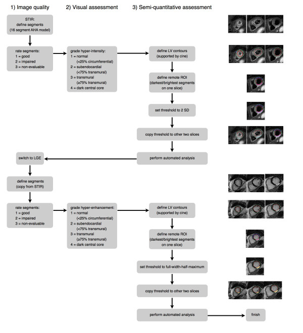
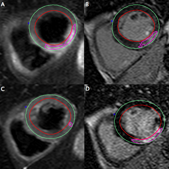
Forty-six consecutive patients with acute myocardial infarction underwent CMR (day 4.5, +/- 1.6) including STIR for the assessment of myocardial edema and late gadolinium enhancement (LGE) for quantification of myocardial necrosis. Thirty of these patients underwent a follow-up CMR at approximately six months (195 +/- 39 days). Both STIR and LGE images were evaluated separately on a segmental basis for image quality as well as for presence and extent of myocardial hyper-intensity, with both visual and semi-quantitative (threshold-based) analysis. LGE was used as a reference standard for localization and extent of myocardial necrosis (acute) or scar (chronic).

RESULTS

Image quality of STIR images was rated as diagnostic in 99.5% of cases. At the acute stage, the sensitivity and specificity of STIR to detect infarcted segments on visual assessment was 95% and 78% respectively, and on semi-quantitative assessment was 99% and 83%, respectively. STIR differentiated acutely from chronically infarcted segments with a sensitivity of 95% by both methods and with a specificity of 99% by visual assessment and 97% by semi-quantitative assessment. The extent of hyper-intense areas on acute STIR images was 85% larger than those on LGE images, with a larger myocardial salvage index in reperfused than in non-reperfused infarcts (p = 0.035).

CONCLUSIONS

STIR with appropriate pulse sequence settings is accurate in detecting acute myocardial infarction (MI) and distinguishing acute from chronic MI with both visual and semi-quantitative analysis. Due to its unique technical characteristics, STIR should be regarded as an edema-weighted rather than a purely T2-weighted technique.

摘要

背景

短反转时间反转恢复(STIR)黑血技术已被用于可视化心肌水肿,从而区分急性和慢性心肌病变。然而,一些心血管磁共振(CMR)组报告了可变的图像质量,因此 STIR 在常规临床实践中的诊断价值受到了质疑。我们的研究目的是分析使用专门用于水肿检测的一组脉冲序列参数的 STIR 的图像质量和诊断性能,并讨论可能影响图像质量的因素。我们假设 STIR 成像在急性心肌梗死的非选择性患者中是一种准确且可靠的检测心肌水肿的方法。

方法

46 例连续的急性心肌梗死患者接受 CMR(第 4.5 天,+/-1.6 天)检查,包括 STIR 以评估心肌水肿和晚期钆增强(LGE)以定量评估心肌坏死。其中 30 例患者在大约 6 个月(195+/-39 天)时进行了随访 CMR。STIR 和 LGE 图像均分别进行节段性评估,评估图像质量以及心肌高信号的存在和程度,包括视觉评估和半定量(基于阈值)分析。LGE 被用作心肌坏死(急性)或瘢痕(慢性)定位和程度的参考标准。

结果

STIR 图像的质量在 99.5%的病例中被评为诊断性。在急性期,STIR 在视觉评估上检测梗死节段的敏感性和特异性分别为 95%和 78%,在半定量评估上分别为 99%和 83%。STIR 通过两种方法分别以 95%的敏感性和 99%的特异性和 97%的半定量评估区分急性和慢性梗死节段。急性 STIR 图像上高信号区域的范围比 LGE 图像大 85%,再灌注性梗死与非再灌注性梗死的心肌挽救指数更大(p=0.035)。

结论

使用适当的脉冲序列设置的 STIR 可准确检测急性心肌梗死(MI),并通过视觉和半定量分析区分急性和慢性 MI。由于其独特的技术特点,STIR 应被视为一种水肿加权而非纯 T2 加权技术。

https://cdn.ncbi.nlm.nih.gov/pmc/blobs/aa90/3350411/8c38a6af680c/1532-429X-14-22-5.jpg
https://cdn.ncbi.nlm.nih.gov/pmc/blobs/aa90/3350411/995fcd53be0f/1532-429X-14-22-1.jpg
https://cdn.ncbi.nlm.nih.gov/pmc/blobs/aa90/3350411/d628d10c93ed/1532-429X-14-22-2.jpg
https://cdn.ncbi.nlm.nih.gov/pmc/blobs/aa90/3350411/197bdb2af60d/1532-429X-14-22-3.jpg
https://cdn.ncbi.nlm.nih.gov/pmc/blobs/aa90/3350411/766a6d1c9e5f/1532-429X-14-22-4.jpg
https://cdn.ncbi.nlm.nih.gov/pmc/blobs/aa90/3350411/8c38a6af680c/1532-429X-14-22-5.jpg

相似文献

1
Cardiovascular magnetic resonance of myocardial edema using a short inversion time inversion recovery (STIR) black-blood technique: diagnostic accuracy of visual and semi-quantitative assessment.心肌水肿的心血管磁共振短反转时间反转恢复(STIR)黑血技术:视觉和半定量评估的诊断准确性。
J Cardiovasc Magn Reson. 2012 Mar 28;14(1):22. doi: 10.1186/1532-429X-14-22.
2
Diagnostic performance of dark-blood T2-weighted CMR for evaluation of acute myocardial injury.心脏磁共振黑血 T2 加权成像在评估急性心肌损伤中的诊断性能。
Invest Radiol. 2013 Jan;48(1):24-31. doi: 10.1097/RLI.0b013e3182718672.
3
Assessment of circumferential endocardial extent of myocardial edema and infarction in patients with reperfused acute myocardial infarction: a cardiovascular magnetic resonance study.再灌注急性心肌梗死患者心肌水肿和梗死的圆周心内膜范围评估:一项心血管磁共振研究
Int Heart J. 2014;55(3):234-8. doi: 10.1536/ihj.13-297. Epub 2014 May 12.
4
Validation of contrast enhanced cine steady-state free precession and T2-weighted CMR for assessment of ischemic myocardial area-at-risk in the presence of reperfusion injury.在存在再灌注损伤的情况下,对比增强电影稳态自由进动和T2加权心脏磁共振成像用于评估缺血心肌危险区域的验证。
Int J Cardiovasc Imaging. 2019 Jun;35(6):1039-1045. doi: 10.1007/s10554-019-01569-x. Epub 2019 Mar 9.
5
Non-contrast T1-mapping detects acute myocardial edema with high diagnostic accuracy: a comparison to T2-weighted cardiovascular magnetic resonance.非对比 T1 映射以高诊断准确性检测急性心肌水肿:与 T2 加权心血管磁共振的比较。
J Cardiovasc Magn Reson. 2012 Jun 21;14(1):42. doi: 10.1186/1532-429X-14-42.
6
CMR imaging of edema in myocardial infarction using cine balanced steady-state free precession.使用电影平衡稳态自由进动技术对心肌梗死水肿进行 CMR 成像。
JACC Cardiovasc Imaging. 2011 Dec;4(12):1265-73. doi: 10.1016/j.jcmg.2011.04.024.
7
T(1) mapping for the diagnosis of acute myocarditis using CMR: comparison to T2-weighted and late gadolinium enhanced imaging.CMR 用于诊断急性心肌炎的 T(1) 映射:与 T2 加权和晚期钆增强成像的比较。
JACC Cardiovasc Imaging. 2013 Oct;6(10):1048-1058. doi: 10.1016/j.jcmg.2013.03.008. Epub 2013 Sep 4.
8
Diagnostic Accuracy of 3.0-T Magnetic Resonance T1 and T2 Mapping and T2-Weighted Dark-Blood Imaging for the Infarct-Related Coronary Artery in Non-ST-Segment Elevation Myocardial Infarction.3.0-T磁共振T1和T2映射及T2加权黑血成像对非ST段抬高型心肌梗死梗死相关冠状动脉的诊断准确性
J Am Heart Assoc. 2017 Mar 31;6(4):e004759. doi: 10.1161/JAHA.116.004759.
9
MultiContrast Delayed Enhancement (MCODE) improves detection of subendocardial myocardial infarction by late gadolinium enhancement cardiovascular magnetic resonance: a clinical validation study.多对比剂延迟强化(MCODE)可提高心脏磁共振钆延迟增强技术诊断心内膜下心肌梗死的准确性:一项临床验证研究。
J Cardiovasc Magn Reson. 2012 Nov 30;14(1):83. doi: 10.1186/1532-429X-14-83.
10
Clinical value of dark-blood late gadolinium enhancement cardiovascular magnetic resonance without additional magnetization preparation.无额外磁化准备的黑血晚期钆增强心血管磁共振的临床价值。
J Cardiovasc Magn Reson. 2019 Jul 29;21(1):44. doi: 10.1186/s12968-019-0556-1.

引用本文的文献

1
In Vivo Fluorine Imaging Using 1.5 Tesla MRI for Depiction of Experimental Myocarditis in a Rodent Animal Model.使用1.5特斯拉磁共振成像进行体内氟成像以描绘啮齿动物模型中的实验性心肌炎
Int J Biomed Imaging. 2023 Jul 14;2023:4659041. doi: 10.1155/2023/4659041. eCollection 2023.
2
SCMR expert consensus statement for cardiovascular magnetic resonance of acquired and non-structural pediatric heart disease.SCMR 专家共识声明:获得性和非结构性儿科心脏病的心血管磁共振成像。
J Cardiovasc Magn Reson. 2022 Jul 21;24(1):44. doi: 10.1186/s12968-022-00873-1.
3
Quantitative Assessment of Late Gadolinium Enhancement and Edema at Cardiac Magnetic Resonance in Low-Risk Myocarditis Patients.

本文引用的文献

1
T2-weighted cardiac MR assessment of the myocardial area-at-risk and salvage area in acute reperfused myocardial infarction: comparison of state-of-the-art dark blood and bright blood T2-weighted sequences.急性再灌注心肌梗死中心肌危险区和挽救区的 T2 加权心脏 MRI 评估:最新的黑血和亮血 T2 加权序列比较。
J Magn Reson Imaging. 2012 Feb;35(2):328-39. doi: 10.1002/jmri.22813. Epub 2011 Sep 29.
2
Clinical characteristics and cardiovascular magnetic resonance findings in stress (takotsubo) cardiomyopathy.应激(心碎)性心肌病的临床特征和心血管磁共振成像表现。
JAMA. 2011 Jul 20;306(3):277-86. doi: 10.1001/jama.2011.992.
3
低危心肌炎患者心脏磁共振钆延迟增强及水肿的定量评估。
Tomography. 2022 Apr 1;8(2):974-984. doi: 10.3390/tomography8020078.
4
VE-Cadherin Is Required for Cardiac Lymphatic Maintenance and Signaling.VE-钙黏蛋白对于心脏淋巴管的维持和信号传递是必需的。
Circ Res. 2022 Jan 7;130(1):5-23. doi: 10.1161/CIRCRESAHA.121.318852. Epub 2021 Nov 18.
5
Black-Blood Contrast in Cardiovascular MRI.心血管磁共振中的黑血对比。
J Magn Reson Imaging. 2022 Jan;55(1):61-80. doi: 10.1002/jmri.27399. Epub 2020 Oct 19.
6
Cardiovascular magnetic resonance in immune checkpoint inhibitor-associated myocarditis.免疫检查点抑制剂相关心肌炎的心血管磁共振成像
Eur Heart J. 2020 May 7;41(18):1733-1743. doi: 10.1093/eurheartj/ehaa051.
7
High T2-weighted signal intensity is associated with myocardial deformation in hypertrophic cardiomyopathy.高 T2 加权信号强度与肥厚型心肌病中的心肌变形有关。
Sci Rep. 2019 Feb 25;9(1):2644. doi: 10.1038/s41598-019-39456-z.
8
Comparison of native myocardial T1 and T2 mapping at 1.5T and 3T in healthy volunteers : Reference values and clinical implications.健康志愿者在1.5T和3T场强下的心肌T1和T2值映射比较:参考值及临床意义
Wien Klin Wochenschr. 2019 Apr;131(7-8):143-155. doi: 10.1007/s00508-018-1411-3. Epub 2018 Dec 5.
9
Cardiac sympathetic neuronal damage precedes myocardial fibrosis in patients with Anderson-Fabry disease.在安德森-法布里病患者中,心肌交感神经损伤先于心肌纤维化。
Eur J Nucl Med Mol Imaging. 2017 Dec;44(13):2266-2273. doi: 10.1007/s00259-017-3778-1. Epub 2017 Jul 22.
10
Detecting Acute Myocardial Infarction by Diffusion-Weighted versus T2-Weighted Imaging and Myocardial Necrosis Markers.通过扩散加权成像与T2加权成像及心肌坏死标志物检测急性心肌梗死
Tex Heart Inst J. 2016 Oct 1;43(5):383-391. doi: 10.14503/THIJ-15-5462. eCollection 2016 Oct.
Prognostic value and determinants of a hypointense infarct core in T2-weighted cardiac magnetic resonance in acute reperfused ST-elevation-myocardial infarction.
急性再灌注 ST 段抬高型心肌梗死 T2 加权心脏磁共振低信号梗死核心的预后价值及其决定因素。
Circ Cardiovasc Imaging. 2011 Jul;4(4):354-62. doi: 10.1161/CIRCIMAGING.110.960500. Epub 2011 Apr 25.
4
T(2) -weighted STIR imaging of myocardial edema associated with ischemia-reperfusion injury: the influence of proton density effect on image contrast.T2 加权 STIR 成像在缺血再灌注损伤相关心肌水肿中的应用:质子密度效应对图像对比度的影响。
J Magn Reson Imaging. 2011 Apr;33(4):962-7. doi: 10.1002/jmri.22456.
5
Direct T2 quantification of myocardial edema in acute ischemic injury.直接 T2 定量评估急性缺血性损伤中的心肌水肿。
JACC Cardiovasc Imaging. 2011 Mar;4(3):269-78. doi: 10.1016/j.jcmg.2010.09.023.
6
Molecular imaging: T2-weighted CMR of the area at risk--a risky business?分子成像:风险区域的T2加权心脏磁共振成像——一项有风险的业务?
Nat Rev Cardiol. 2010 Oct;7(10):547-9. doi: 10.1038/nrcardio.2010.124.
7
Myocardial salvage by CMR correlates with LV remodeling and early ST-segment resolution in acute myocardial infarction.CMR 检测的心肌挽救与急性心肌梗死后的 LV 重构和早期 ST 段回落相关。
JACC Cardiovasc Imaging. 2010 Jan;3(1):45-51. doi: 10.1016/j.jcmg.2009.06.016.
8
T2 quantification for improved detection of myocardial edema.用于提高心肌水肿检测的 T2 定量
J Cardiovasc Magn Reson. 2009 Dec 30;11(1):56. doi: 10.1186/1532-429X-11-56.
9
Intramyocardial hemorrhage and microvascular obstruction after primary percutaneous coronary intervention.心肌内出血和经皮冠状动脉介入治疗后的微血管阻塞。
Int J Cardiovasc Imaging. 2010 Jan;26(1):49-55. doi: 10.1007/s10554-009-9499-1. Epub 2009 Sep 15.
10
Myocardium at risk after acute infarction in humans on cardiac magnetic resonance: quantitative assessment during follow-up and validation with single-photon emission computed tomography.人类急性心肌梗死后心脏磁共振成像显示的危险心肌:随访期间的定量评估及与单光子发射计算机断层扫描的验证
JACC Cardiovasc Imaging. 2009 May;2(5):569-76. doi: 10.1016/j.jcmg.2008.11.018.