• 文献检索
  • 文档翻译
  • 深度研究
  • 学术资讯
  • Suppr Zotero 插件Zotero 插件
  • 邀请有礼
  • 套餐&价格
  • 历史记录
应用&插件
Suppr Zotero 插件Zotero 插件浏览器插件Mac 客户端Windows 客户端微信小程序
定价
高级版会员购买积分包购买API积分包
服务
文献检索文档翻译深度研究API 文档MCP 服务
关于我们
关于 Suppr公司介绍联系我们用户协议隐私条款
关注我们

Suppr 超能文献

核心技术专利:CN118964589B侵权必究
粤ICP备2023148730 号-1Suppr @ 2026

文献检索

告别复杂PubMed语法,用中文像聊天一样搜索,搜遍4000万医学文献。AI智能推荐,让科研检索更轻松。

立即免费搜索

文件翻译

保留排版,准确专业,支持PDF/Word/PPT等文件格式,支持 12+语言互译。

免费翻译文档

深度研究

AI帮你快速写综述,25分钟生成高质量综述,智能提取关键信息,辅助科研写作。

立即免费体验

从临床 CT 评估股皮质:厚度、密度和质量。

Imaging the femoral cortex: thickness, density and mass from clinical CT.

机构信息

University of Cambridge, Department of Engineering, Trumpington Street, Cambridge, UK.

出版信息

Med Image Anal. 2012 Jul;16(5):952-65. doi: 10.1016/j.media.2012.02.008. Epub 2012 Feb 28.

DOI:10.1016/j.media.2012.02.008
PMID:22465079
原文链接:https://pmc.ncbi.nlm.nih.gov/articles/PMC3417239/
Abstract

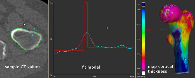
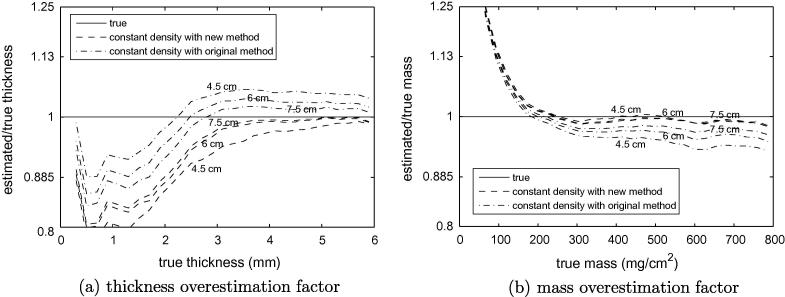
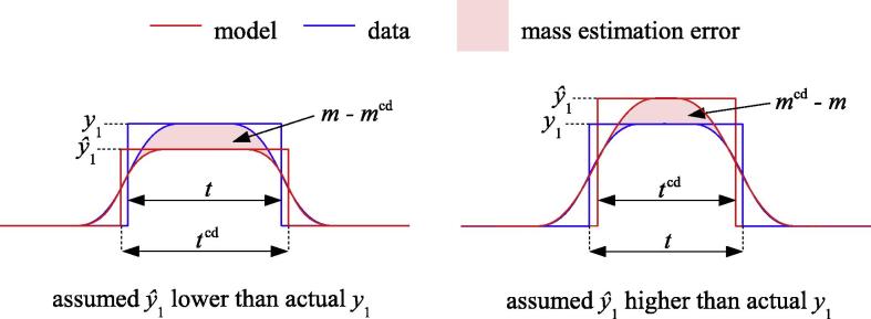
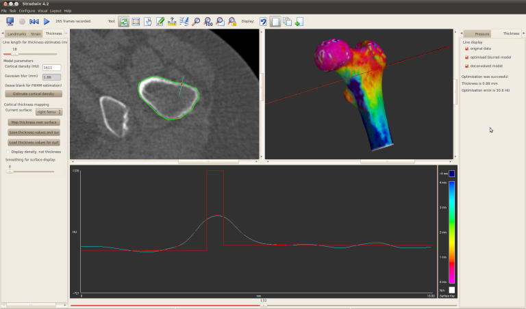
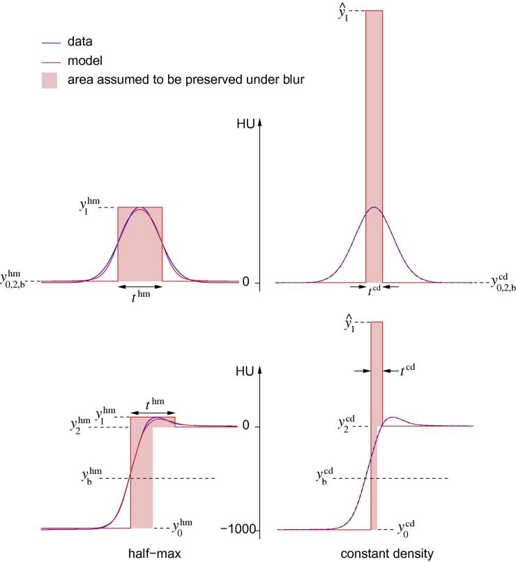
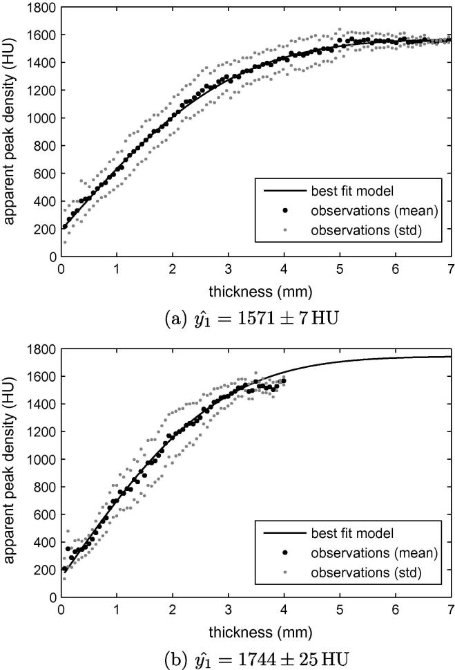
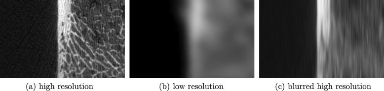
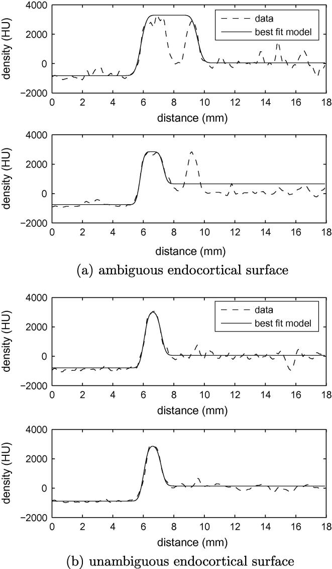
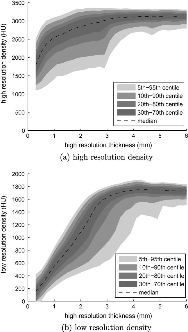
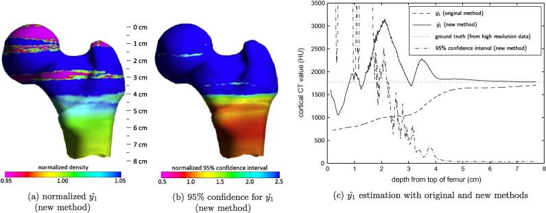
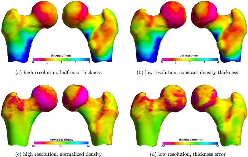
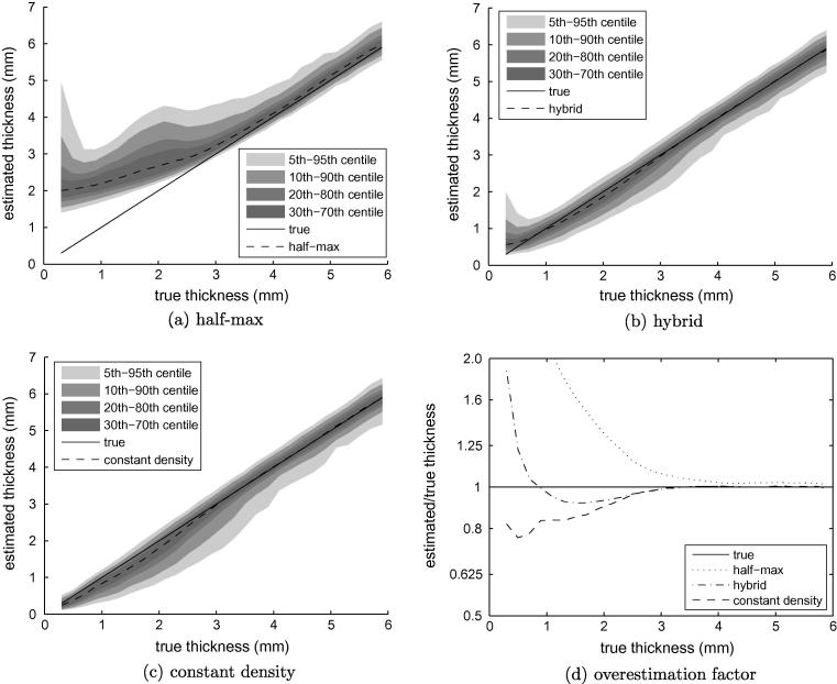
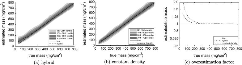
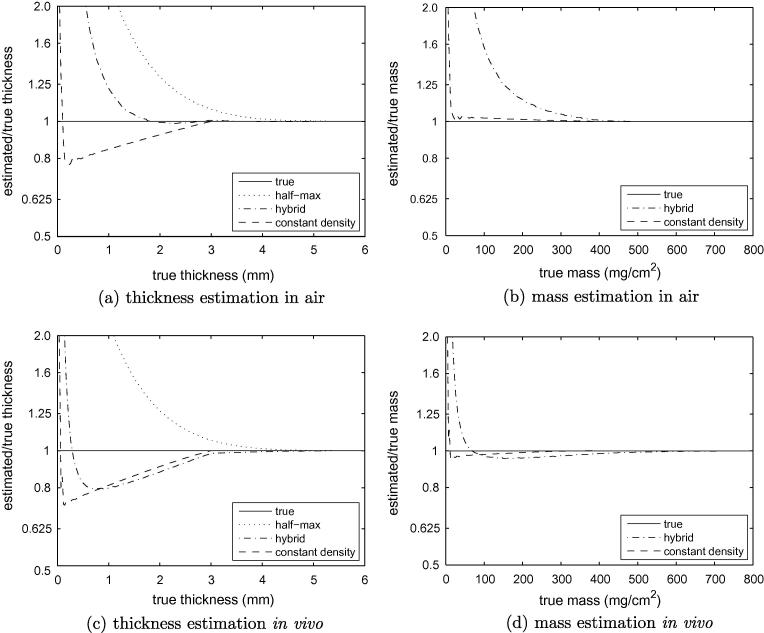
There is growing evidence that focal thinning of cortical bone in the proximal femur may predispose a hip to fracture. Detecting such defects in clinical CT is challenging, since cortices may be significantly thinner than the imaging system's point spread function. We recently proposed a model-fitting technique to measure sub-millimetre cortices, an ill-posed problem which was regularized by assuming a specific, fixed value for the cortical density. In this paper, we develop the work further by proposing and evaluating a more rigorous method for estimating the constant cortical density, and extend the paradigm to encompass the mapping of cortical mass (mineral mg/cm(2)) in addition to thickness. Density, thickness and mass estimates are evaluated on sixteen cadaveric femurs, with high resolution measurements from a micro-CT scanner providing the gold standard. The results demonstrate robust, accurate measurement of peak cortical density and cortical mass. Cortical thickness errors are confined to regions of thin cortex and are bounded by the extent to which the local density deviates from the peak, averaging 20% for 0.5mm cortex.

摘要

越来越多的证据表明,股骨近端皮质骨的局灶性变薄可能使髋关节容易发生骨折。在临床 CT 中检测到这种缺陷具有挑战性,因为皮质可能比成像系统的点扩散函数薄得多。我们最近提出了一种模型拟合技术来测量亚毫米皮质,这是一个不适定的问题,通过假设皮质密度的特定固定值来正则化。在本文中,我们通过提出和评估一种更严格的方法来估计固定皮质密度来进一步开展这项工作,并将范例扩展到除厚度之外还包括皮质质量(mg/cm^2 矿物质)的映射。使用高分辨率的微 CT 扫描仪进行的十六具尸体股骨的测量对密度、厚度和质量估计值进行了评估,为金标准提供了参考。结果表明,皮质的峰值密度和皮质质量的测量是稳健和准确的。皮质厚度的误差局限于皮质较薄的区域,并且受到局部密度偏离峰值的程度的限制,平均为 0.5mm 皮质的 20%。

https://cdn.ncbi.nlm.nih.gov/pmc/blobs/1c9c/3417239/119a88fd16e0/gr13.jpg
https://cdn.ncbi.nlm.nih.gov/pmc/blobs/1c9c/3417239/ce7653cd23c7/fx1.jpg
https://cdn.ncbi.nlm.nih.gov/pmc/blobs/1c9c/3417239/3950dceac19d/gr14.jpg
https://cdn.ncbi.nlm.nih.gov/pmc/blobs/1c9c/3417239/cc4546bb1cfd/gr15.jpg
https://cdn.ncbi.nlm.nih.gov/pmc/blobs/1c9c/3417239/b791dedb438e/gr1.jpg
https://cdn.ncbi.nlm.nih.gov/pmc/blobs/1c9c/3417239/73aa89b6c84b/gr2.jpg
https://cdn.ncbi.nlm.nih.gov/pmc/blobs/1c9c/3417239/3c2380baa70f/gr3.jpg
https://cdn.ncbi.nlm.nih.gov/pmc/blobs/1c9c/3417239/1861bc6822be/gr4.jpg
https://cdn.ncbi.nlm.nih.gov/pmc/blobs/1c9c/3417239/432de1fdbfaa/gr5.jpg
https://cdn.ncbi.nlm.nih.gov/pmc/blobs/1c9c/3417239/8bea878c1679/gr6.jpg
https://cdn.ncbi.nlm.nih.gov/pmc/blobs/1c9c/3417239/3e2d6f2fd602/gr7.jpg
https://cdn.ncbi.nlm.nih.gov/pmc/blobs/1c9c/3417239/2b50be9c8786/gr8.jpg
https://cdn.ncbi.nlm.nih.gov/pmc/blobs/1c9c/3417239/afe81bedf248/gr9.jpg
https://cdn.ncbi.nlm.nih.gov/pmc/blobs/1c9c/3417239/1af8349ccf27/gr10.jpg
https://cdn.ncbi.nlm.nih.gov/pmc/blobs/1c9c/3417239/b798ce56ba55/gr11.jpg
https://cdn.ncbi.nlm.nih.gov/pmc/blobs/1c9c/3417239/a86aff360c81/gr12.jpg
https://cdn.ncbi.nlm.nih.gov/pmc/blobs/1c9c/3417239/119a88fd16e0/gr13.jpg

相似文献

1
Imaging the femoral cortex: thickness, density and mass from clinical CT.从临床 CT 评估股皮质:厚度、密度和质量。
Med Image Anal. 2012 Jul;16(5):952-65. doi: 10.1016/j.media.2012.02.008. Epub 2012 Feb 28.
2
Generation of 3D shape, density, cortical thickness and finite element mesh of proximal femur from a DXA image.从 DXA 图像生成近端股骨的 3D 形状、密度、皮质厚度和有限元网格。
Med Image Anal. 2015 Aug;24(1):125-134. doi: 10.1016/j.media.2015.06.001. Epub 2015 Jun 19.
3
High resolution cortical bone thickness measurement from clinical CT data.从临床 CT 数据中测量高分辨率皮质骨厚度。
Med Image Anal. 2010 Jun;14(3):276-90. doi: 10.1016/j.media.2010.01.003. Epub 2010 Jan 25.
4
Measurement of the bone endocortical region using clinical CT.临床 CT 测量骨内皮质区
Med Image Anal. 2018 Feb;44:28-40. doi: 10.1016/j.media.2017.11.006. Epub 2017 Nov 16.
5
Independent measurement of femoral cortical thickness and cortical bone density using clinical CT.临床 CT 独立测量股骨皮质厚度和皮质骨密度。
Med Image Anal. 2015 Feb;20(1):249-64. doi: 10.1016/j.media.2014.11.012. Epub 2014 Dec 5.
6
Technical Note: Cortical thickness and density estimation from clinical CT using a prior thickness-density relationship.技术说明:利用先前的厚度-密度关系从临床CT估计皮质厚度和密度
Med Phys. 2016 Apr;43(4):1945. doi: 10.1118/1.4944501.
7
Distribution of bone density and cortical thickness in the proximal femur and their association with hip fracture in postmenopausal women: a quantitative computed tomography study.绝经后女性股骨近端骨密度和皮质厚度的分布及其与髋部骨折的关系:一项定量 CT 研究。
Osteoporos Int. 2014 Jan;25(1):251-63. doi: 10.1007/s00198-013-2401-y. Epub 2013 May 30.
8
Bone turnover markers are associated with higher cortical porosity, thinner cortices, and larger size of the proximal femur and non-vertebral fractures.骨转换标志物与较高的皮质骨孔隙率、较薄的皮质骨以及股骨近端较大的尺寸和非椎体骨折相关。
Bone. 2015 Dec;81:1-6. doi: 10.1016/j.bone.2015.06.016. Epub 2015 Jun 22.
9
Which Two-dimensional Radiographic Measurements of Cam Femoroacetabular Impingement Best Describe the Three-dimensional Shape of the Proximal Femur?Cam 型股骨髋臼撞击症的二维放射学测量中,哪些指标最能描述股骨近端的三维形态?
Clin Orthop Relat Res. 2019 Jan;477(1):242-253. doi: 10.1097/CORR.0000000000000462.
10
Cortical and Trabecular Bone Analysis of Patients With High Bone Mass From the Barcelona Osteoporosis Cohort Using 3-Dimensional Dual-Energy X-ray Absorptiometry: A Case-Control Study.使用三维双能 X 射线吸收法对巴塞罗那骨质疏松队列中高骨量患者的皮质骨和小梁骨进行分析:一项病例对照研究。
J Clin Densitom. 2018 Oct-Dec;21(4):480-484. doi: 10.1016/j.jocd.2017.05.012. Epub 2017 Jun 22.

引用本文的文献

1
How effective is proximal fibular osteotomy in redistributing joint pressures? Insights from an HTO comparative in-silico study.腓骨近端截骨术在重新分配关节压力方面的效果如何?来自 HTO 比较的计算机模拟研究的见解。
J Orthop Surg Res. 2024 Jun 4;19(1):333. doi: 10.1186/s13018-024-04807-8.
2
Experimental study on determining the degree of bone healing by wall thickness ratio analysis.通过骨壁厚度比分析来确定骨愈合程度的实验研究。
J Orthop Surg Res. 2024 Jan 19;19(1):79. doi: 10.1186/s13018-024-04565-7.
3
A Correspondence-Based Network Approach for Groupwise Analysis of Patient-Specific Spatiotemporal Data.

本文引用的文献

1
Targeted regeneration of bone in the osteoporotic human femur.靶向修复骨质疏松症患者股骨的骨。
PLoS One. 2011 Jan 14;6(1):e16190. doi: 10.1371/journal.pone.0016190.
2
High resolution cortical bone thickness measurement from clinical CT data.从临床 CT 数据中测量高分辨率皮质骨厚度。
Med Image Anal. 2010 Jun;14(3):276-90. doi: 10.1016/j.media.2010.01.003. Epub 2010 Jan 25.
3
Changing structure of the femoral neck across the adult female lifespan.成年女性一生中股骨颈结构的变化。
基于对应网络的方法用于分析特定患者的时空数据。
Ann Biomed Eng. 2023 Oct;51(10):2289-2300. doi: 10.1007/s10439-023-03270-6. Epub 2023 Jun 25.
4
Micron-resolution Imaging of Cortical Bone under 14 T Ultrahigh Magnetic Field.在 14T 超高磁场下对皮质骨进行微米分辨率成像。
Adv Sci (Weinh). 2023 Aug;10(24):e2300959. doi: 10.1002/advs.202300959. Epub 2023 Jun 20.
5
Advancing Osteoporosis Evaluation Procedures: Detailed Computational Analysis of Regional Structural Vulnerabilities in Osteoporotic Bone.推进骨质疏松症评估程序:骨质疏松性骨区域结构脆弱性的详细计算分析
J Pers Med. 2023 Feb 13;13(2):321. doi: 10.3390/jpm13020321.
6
Automated volumetric and statistical shape assessment of cam-type morphology of the femoral head-neck region from clinical 3D magnetic resonance images.基于临床三维磁共振图像的股骨头颈区域凸轮型形态的自动容积和统计形状评估
Quant Imaging Med Surg. 2022 Oct;12(10):4924-4941. doi: 10.21037/qims-22-332.
7
The West China Hospital radiographic classification for fibrous dysplasia in femur and adjacent bones: A retrospective analysis of 205 patients.华西医院股骨及相邻骨纤维结构不良的影像学分类:205 例回顾性分析。
Orthop Surg. 2022 Sep;14(9):2096-2108. doi: 10.1111/os.13404. Epub 2022 Aug 4.
8
Cetaceans Humerus Radiodensity by CT: A Useful Technique Differentiating between Species, Ecophysiology, and Age.通过CT检测鲸类肱骨的骨密度:一种区分物种、生态生理学特征和年龄的有用技术。
Animals (Basel). 2022 Jul 13;12(14):1793. doi: 10.3390/ani12141793.
9
Protective effect of bisphosphonate on the cortical bone at key locations of the femur in aromatase inhibitor-associated bone loss: A three-dimensional cortical bone mapping study.双膦酸盐对芳香化酶抑制剂相关性骨质流失时股骨关键部位皮质骨的保护作用:一项三维皮质骨图谱研究。
J Bone Oncol. 2021 Dec 29;32:100409. doi: 10.1016/j.jbo.2021.100409. eCollection 2022 Feb.
10
Spatial distribution of hip cortical thickness in postmenopausal women with different osteoporotic fractures.绝经后不同骨质疏松性骨折女性髋部皮质骨厚度的空间分布。
Arch Osteoporos. 2021 Nov 15;16(1):172. doi: 10.1007/s11657-021-01039-9.
J Bone Miner Res. 2010 Mar;25(3):482-91. doi: 10.1359/jbmr.090734.
4
During sideways falls proximal femur fractures initiate in the superolateral cortex: evidence from high-speed video of simulated fractures.在侧方跌倒时,股骨近端骨折始于上外侧皮质:来自模拟骨折高速视频的证据。
J Biomech. 2009 Aug 25;42(12):1917-25. doi: 10.1016/j.jbiomech.2009.05.001. Epub 2009 Jun 13.
5
Hip fractures and the contribution of cortical versus trabecular bone to femoral neck strength.髋部骨折以及皮质骨与小梁骨对股骨颈强度的贡献。
J Bone Miner Res. 2009 Mar;24(3):468-74. doi: 10.1359/jbmr.081108.
6
Prediction of incident hip fracture risk by femur geometry variables measured by hip structural analysis in the study of osteoporotic fractures.在骨质疏松性骨折研究中,通过髋部结构分析测量的股骨几何变量预测髋部骨折发生风险。
J Bone Miner Res. 2008 Dec;23(12):1892-904. doi: 10.1359/jbmr.080802.
7
Considerations for development of surrogate endpoints for antifracture efficacy of new treatments in osteoporosis: a perspective.骨质疏松症新疗法抗骨折疗效替代终点指标开发的考量:一种观点
J Bone Miner Res. 2008 Aug;23(8):1155-67. doi: 10.1359/jbmr.080301.
8
European guidance for the diagnosis and management of osteoporosis in postmenopausal women.欧洲绝经后女性骨质疏松症诊断与管理指南。
Osteoporos Int. 2008 Apr;19(4):399-428. doi: 10.1007/s00198-008-0560-z. Epub 2008 Feb 12.
9
Load distribution in the healthy and osteoporotic human proximal femur during a fall to the side.向一侧摔倒时健康及骨质疏松的人体近端股骨的负荷分布
Bone. 2008 Jan;42(1):30-5. doi: 10.1016/j.bone.2007.08.039. Epub 2007 Sep 6.
10
Model-based cartilage thickness measurement in the submillimeter range.基于模型的亚毫米范围内软骨厚度测量。
Med Phys. 2007 Sep;34(9):3562-70. doi: 10.1118/1.2766759.