Department of Radiation Oncology, Washington University School of Medicine, St Louis, MO 63110-6311, USA.
Int J Radiat Oncol Biol Phys. 2012 Jul 1;83(3):e353-62. doi: 10.1016/j.ijrobp.2012.01.023. Epub 2012 Apr 6.
To define a male and female pelvic normal tissue contouring atlas for Radiation Therapy Oncology Group (RTOG) trials.
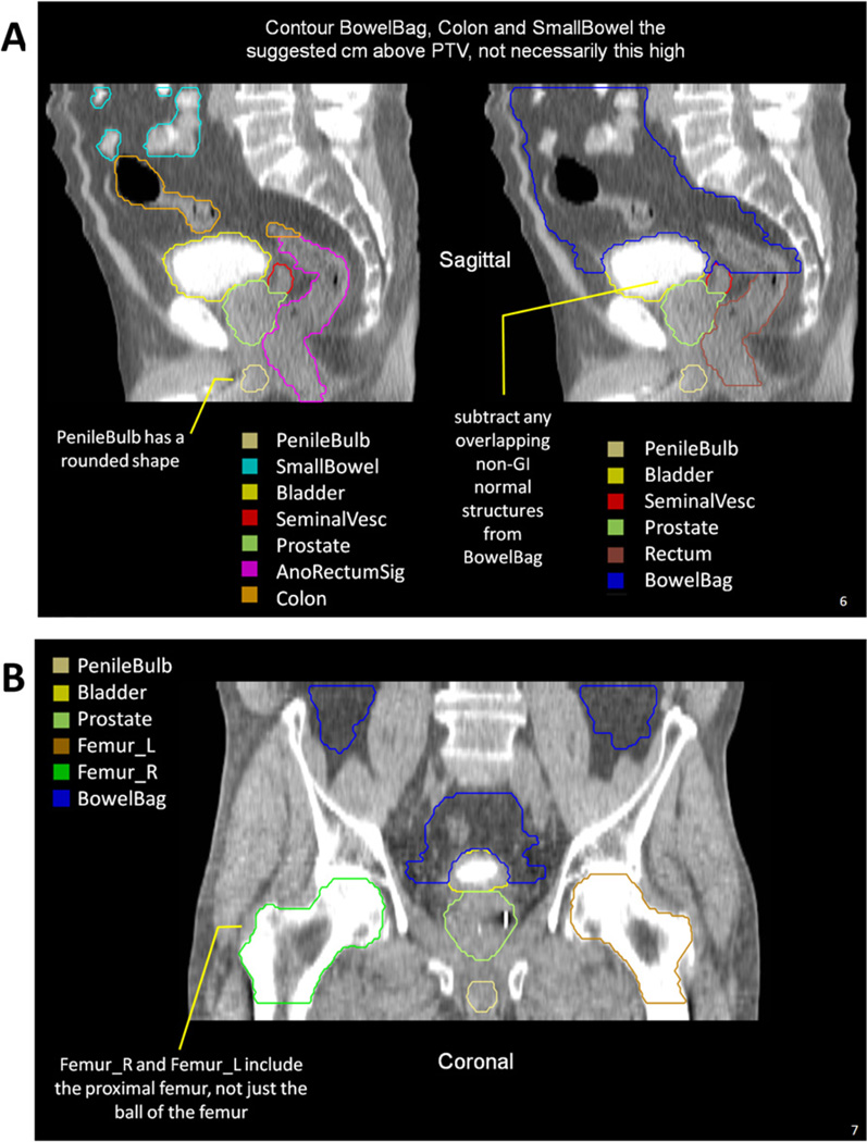
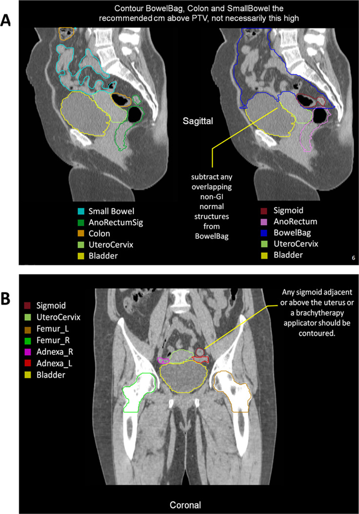
One male pelvis computed tomography (CT) data set and one female pelvis CT data set were shared via the Image-Guided Therapy QA Center. A total of 16 radiation oncologists participated. The following organs at risk were contoured in both CT sets: anus, anorectum, rectum (gastrointestinal and genitourinary definitions), bowel NOS (not otherwise specified), small bowel, large bowel, and proximal femurs. The following were contoured in the male set only: bladder, prostate, seminal vesicles, and penile bulb. The following were contoured in the female set only: uterus, cervix, and ovaries. A computer program used the binomial distribution to generate 95% group consensus contours. These contours and definitions were then reviewed by the group and modified.
The panel achieved consensus definitions for pelvic normal tissue contouring in RTOG trials with these standardized names: Rectum, AnoRectum, SmallBowel, Colon, BowelBag, Bladder, UteroCervix, Adnexa_R, Adnexa_L, Prostate, SeminalVesc, PenileBulb, Femur_R, and Femur_L. Two additional normal structures whose purpose is to serve as targets in anal and rectal cancer were defined: AnoRectumSig and Mesorectum. Detailed target volume contouring guidelines and images are discussed.
Consensus guidelines for pelvic normal tissue contouring were reached and are available as a CT image atlas on the RTOG Web site. This will allow uniformity in defining normal tissues for clinical trials delivering pelvic radiation and will facilitate future normal tissue complication research.
为放射治疗肿瘤学组(RTOG)试验定义男性和女性骨盆正常组织轮廓图谱。
通过图像引导治疗质量保证中心共享了一个男性骨盆 CT 数据集和一个女性骨盆 CT 数据集。共有 16 名放射肿瘤学家参与。在这两个 CT 数据集上,对以下危及器官进行了轮廓勾画:肛门、肛门直肠、直肠(胃肠和泌尿生殖系统定义)、肠道 NOS(其他未指定)、小肠、大肠和股骨近端。仅在男性数据集上对以下器官进行了轮廓勾画:膀胱、前列腺、精囊和阴茎球。仅在女性数据集上对以下器官进行了轮廓勾画:子宫、宫颈和卵巢。使用二项式分布的计算机程序生成了 95%的组共识轮廓。然后,专家组对这些轮廓和定义进行了审查和修改。
专家组就 RTOG 试验中骨盆正常组织轮廓勾画达成了共识定义,使用了这些标准化名称:直肠、肛门直肠、小肠、结肠、肠道袋、膀胱、子宫颈-子宫体、附件 R、附件 L、前列腺、精囊、阴茎球、股骨 R 和股骨 L。还定义了两个用于肛门和直肠癌症的额外正常结构作为靶区:肛门直肠标志和直肠系膜。详细的靶区勾画指南和图像进行了讨论。
达成了骨盆正常组织轮廓勾画的共识指南,并作为 RTOG 网站上的 CT 图像图谱提供。这将允许在进行骨盆放射治疗的临床试验中对正常组织进行统一定义,并为未来的正常组织并发症研究提供便利。