Liebscher Sabine, Meyer-Luehmann Melanie
Department of Cellular and Systems Neurobiology, Max Planck Institute of Neurobiology Martinsried, Germany.
Front Psychiatry. 2012 Apr 2;3:26. doi: 10.3389/fpsyt.2012.00026. eCollection 2012.
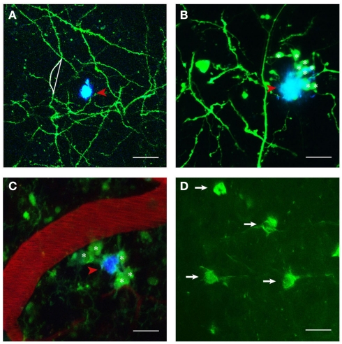
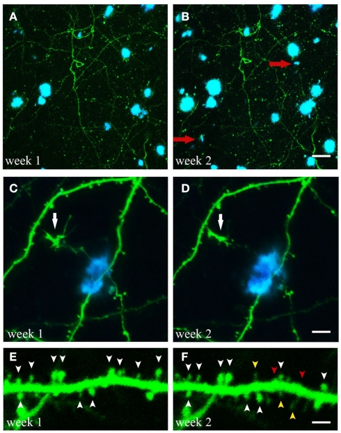
Alzheimer's disease (AD) is a protein conformational disorder characterized by two major neuropathological features: extracellular accumulations of amyloid-β peptides in the form of plaques and intracellular tangles, consisting of hyperphosphorylated tau proteins. Several morphological and functional changes are associated with these lesions in the diseased brain, such as dendritic and synaptic alterations, as well as microglial and astroglial recruitment and their activation. The availability of transgenic mouse models that mimic key aspects of the disease in conjunction with recent advances in two-photon imaging facilitate the study of fundamental aspects of AD pathogenesis and allow for longitudinally monitoring the efficacy of therapeutic interventions. Here, we review the ambitious efforts to understand the relationship between the main neuropathological hallmarks of AD and their associated structural and functional abnormalities by means of in vivo two-photon imaging.
阿尔茨海默病(AD)是一种蛋白质构象紊乱疾病,其特征为两个主要的神经病理学特征:以斑块形式存在的细胞外淀粉样β肽聚集以及由高度磷酸化的tau蛋白组成的细胞内缠结。在患病大脑中,这些病变会伴随一些形态和功能变化,如树突和突触改变,以及小胶质细胞和星形胶质细胞的募集及其激活。转基因小鼠模型可模拟该疾病的关键特征,结合双光子成像技术的最新进展,有助于研究AD发病机制的基本方面,并能够纵向监测治疗干预的效果。在此,我们回顾了通过体内双光子成像来理解AD主要神经病理学特征与其相关结构和功能异常之间关系的宏大努力。