• 文献检索
  • 文档翻译
  • 深度研究
  • 学术资讯
  • Suppr Zotero 插件Zotero 插件
  • 邀请有礼
  • 套餐&价格
  • 历史记录
应用&插件
Suppr Zotero 插件Zotero 插件浏览器插件Mac 客户端Windows 客户端微信小程序
定价
高级版会员购买积分包购买API积分包
服务
文献检索文档翻译深度研究API 文档MCP 服务
关于我们
关于 Suppr公司介绍联系我们用户协议隐私条款
关注我们

Suppr 超能文献

核心技术专利:CN118964589B侵权必究
粤ICP备2023148730 号-1Suppr @ 2026

文献检索

告别复杂PubMed语法,用中文像聊天一样搜索,搜遍4000万医学文献。AI智能推荐,让科研检索更轻松。

立即免费搜索

文件翻译

保留排版,准确专业,支持PDF/Word/PPT等文件格式,支持 12+语言互译。

免费翻译文档

深度研究

AI帮你快速写综述,25分钟生成高质量综述,智能提取关键信息,辅助科研写作。

立即免费体验

利用受激拉曼散射显微镜对阿尔茨海默病中的淀粉样斑块进行无标记成像。

Label-free imaging of amyloid plaques in Alzheimer's disease with stimulated Raman scattering microscopy.

机构信息

State Key Laboratory of Surface Physics and Department of Physics, Multiscale Research Institute of Complex Systems, Human Phenome Institute, Key Laboratory of Micro and Nano Photonics Structures (Ministry of Education), Fudan University, Shanghai 200433, China.

Department of Chemistry and Chemical Biology, Harvard University, Cambridge, MA 02138, USA.

出版信息

Sci Adv. 2018 Nov 16;4(11):eaat7715. doi: 10.1126/sciadv.aat7715. eCollection 2018 Nov.

DOI:10.1126/sciadv.aat7715
PMID:30456301
原文链接:https://pmc.ncbi.nlm.nih.gov/articles/PMC6239428/
Abstract

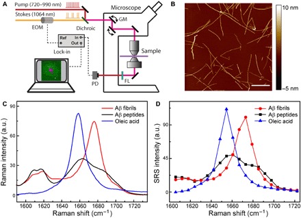
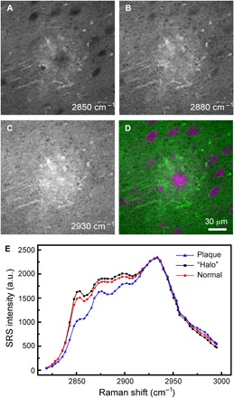
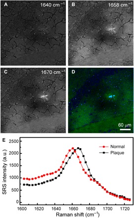
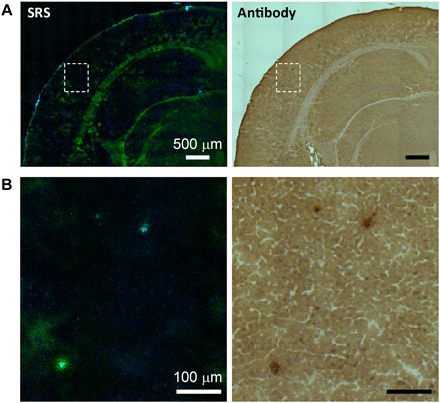
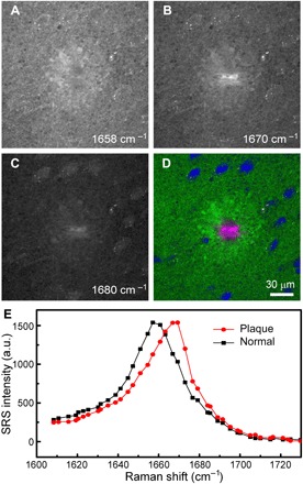
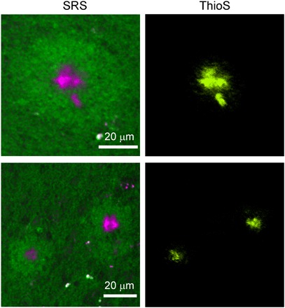
One of the key pathological features of Alzheimer's disease (AD) is the existence of extracellular deposition of amyloid plaques formed with misfolded amyloid-β (Aβ). The conformational change of proteins leads to enriched contents of β sheets, resulting in remarkable changes of vibrational spectra, especially the spectral shifts of the amide I mode. Here, we applied stimulated Raman scattering (SRS) microscopy to image amyloid plaques in the brain tissue of an AD mouse model. We have demonstrated the capability of SRS microscopy as a rapid, label-free imaging modality to differentiate misfolded from normal proteins based on the blue shift (~10 cm) of amide I SRS spectra. Furthermore, SRS imaging of Aβ plaques was verified by antibody staining of frozen thin sections and fluorescence imaging of fresh tissues. Our method may provide a new approach for studies of AD pathology, as well as other neurodegenerative diseases associated with protein misfolding.

摘要

阿尔茨海默病(AD)的一个关键病理学特征是存在由错误折叠的淀粉样-β(Aβ)形成的细胞外淀粉样斑块沉积。蛋白质构象的改变导致β片层含量增加,从而导致振动光谱发生显著变化,特别是酰胺 I 模式的光谱位移。在这里,我们应用受激拉曼散射(SRS)显微镜来对 AD 小鼠模型脑组织中的淀粉样斑块进行成像。我们已经证明了 SRS 显微镜作为一种快速、无标记的成像方式的能力,它可以根据酰胺 I SRS 光谱的蓝移(约 10 cm)来区分错误折叠的蛋白质与正常蛋白质。此外,我们通过对冷冻薄片进行抗体染色和对新鲜组织进行荧光成像来验证了 Aβ斑块的 SRS 成像。我们的方法可能为 AD 病理学以及其他与蛋白质错误折叠相关的神经退行性疾病的研究提供新的途径。

https://cdn.ncbi.nlm.nih.gov/pmc/blobs/2f5a/6239428/be734e488242/aat7715-F6.jpg
https://cdn.ncbi.nlm.nih.gov/pmc/blobs/2f5a/6239428/6605aaa02d0c/aat7715-F1.jpg
https://cdn.ncbi.nlm.nih.gov/pmc/blobs/2f5a/6239428/777f889dcb7c/aat7715-F2.jpg
https://cdn.ncbi.nlm.nih.gov/pmc/blobs/2f5a/6239428/5c8245e4cb3b/aat7715-F3.jpg
https://cdn.ncbi.nlm.nih.gov/pmc/blobs/2f5a/6239428/2f8dca20b397/aat7715-F4.jpg
https://cdn.ncbi.nlm.nih.gov/pmc/blobs/2f5a/6239428/64b633792c67/aat7715-F5.jpg
https://cdn.ncbi.nlm.nih.gov/pmc/blobs/2f5a/6239428/be734e488242/aat7715-F6.jpg

相似文献

1
Label-free imaging of amyloid plaques in Alzheimer's disease with stimulated Raman scattering microscopy.利用受激拉曼散射显微镜对阿尔茨海默病中的淀粉样斑块进行无标记成像。
Sci Adv. 2018 Nov 16;4(11):eaat7715. doi: 10.1126/sciadv.aat7715. eCollection 2018 Nov.
2
Nonlinear and vibrational microscopy for label-free characterization of amyloid-β plaques in Alzheimer's disease model.用于阿尔茨海默病模型中无标记淀粉样β斑块特征分析的非线性和振动显微镜。
Analyst. 2021 May 4;146(9):2945-2954. doi: 10.1039/d1an00074h.
3
Label-free vibrational imaging of different Aβ plaque types in Alzheimer's disease reveals sequential events in plaque development.无标记振动成像技术对阿尔茨海默病中不同 Aβ 斑块类型的研究揭示了斑块发展过程中的顺序事件。
Acta Neuropathol Commun. 2020 Dec 11;8(1):222. doi: 10.1186/s40478-020-01091-5.
4
Multimodal, label-free fluorescence and Raman imaging of amyloid deposits in snap-frozen Alzheimer's disease human brain tissue.对阿尔茨海默病患者脑速冻组织中淀粉样蛋白沉积物的多模态、无标记荧光和拉曼成像。
Commun Biol. 2021 Apr 15;4(1):474. doi: 10.1038/s42003-021-01981-x.
5
Probing amyloid-β pathology in transgenic Alzheimer's disease (tgArcSwe) mice using MALDI imaging mass spectrometry.使用基质辅助激光解吸电离成像质谱法探究转基因阿尔茨海默病(tgArcSwe)小鼠中的淀粉样β病理。
J Neurochem. 2016 Aug;138(3):469-78. doi: 10.1111/jnc.13645. Epub 2016 May 26.
6
Microglia contributes to plaque growth by cell death due to uptake of amyloid β in the brain of Alzheimer's disease mouse model.在阿尔茨海默病小鼠模型的大脑中,小胶质细胞因摄取β淀粉样蛋白而导致细胞死亡,从而促进斑块生长。
Glia. 2016 Dec;64(12):2274-2290. doi: 10.1002/glia.23074. Epub 2016 Sep 23.
7
Human Alzheimer's disease gene expression signatures and immune profile in APP mouse models: a discrete transcriptomic view of Aβ plaque pathology.人阿尔茨海默病基因表达特征和 APP 小鼠模型中的免疫特征:Aβ 斑块病理学的离散转录组学观点。
J Neuroinflammation. 2018 Sep 6;15(1):256. doi: 10.1186/s12974-018-1265-7.
8
Shedding Light on the Molecular Pathology of Amyloid Plaques in Transgenic Alzheimer's Disease Mice Using Multimodal MALDI Imaging Mass Spectrometry.利用多模态 MALDI 成像质谱法揭示转基因阿尔茨海默病小鼠中淀粉样斑块的分子病理学。
ACS Chem Neurosci. 2018 Jul 18;9(7):1802-1817. doi: 10.1021/acschemneuro.8b00121. Epub 2018 May 4.
9
Distinguishing Amyloid β-Protein in a Mouse Model of Alzheimer's Disease by Label-Free Vibrational Imaging.通过无标记振动成像在阿尔茨海默病的小鼠模型中区分淀粉样β-蛋白。
Biosensors (Basel). 2021 Sep 30;11(10):365. doi: 10.3390/bios11100365.
10
Intraneuronal Aβ accumulation, amyloid plaques, and synapse pathology in Alzheimer's disease.阿尔茨海默病中的细胞内 Aβ 积累、淀粉样斑块和突触病变。
Neurodegener Dis. 2012;10(1-4):56-9. doi: 10.1159/000334762. Epub 2012 Jan 21.

引用本文的文献

1
Functional Raman Probes for Detecting Enzyme Activities Based on Aggregation Control.基于聚集控制的用于检测酶活性的功能性拉曼探针。
Anal Chem. 2025 Sep 9;97(35):19057-19065. doi: 10.1021/acs.analchem.5c02231. Epub 2025 Aug 27.
2
Shadowless amyloid imaging with quantitative birefringence contrast.具有定量双折射对比度的无阴影淀粉样蛋白成像。
Sci Rep. 2025 Aug 6;15(1):28776. doi: 10.1038/s41598-025-11795-0.
3
Low-frequency Raman spectra of amyloid fibrils.淀粉样纤维的低频拉曼光谱。

本文引用的文献

1
Hyperspectral Raman imaging of neuritic plaques and neurofibrillary tangles in brain tissue from Alzheimer's disease patients.阿尔茨海默病患者脑组织中神经原纤维缠结和神经纤维缠结的高光谱拉曼成像。
Sci Rep. 2017 Nov 15;7(1):15603. doi: 10.1038/s41598-017-16002-3.
2
Structural Changes of Amyloid Beta in Hippocampus of Rats Exposed to Ozone: A Raman Spectroscopy Study.暴露于臭氧环境的大鼠海马中β淀粉样蛋白的结构变化:一项拉曼光谱研究
Front Mol Neurosci. 2017 May 22;10:137. doi: 10.3389/fnmol.2017.00137. eCollection 2017.
3
Stimulated Raman scattering microscopy and spectroscopy with a rapid scanning optical delay line.
J Chem Phys. 2025 Jun 7;162(21). doi: 10.1063/5.0260500.
4
Chemical imaging for biological systems: techniques, AI-driven processing, and applications.生物系统的化学成像:技术、人工智能驱动的处理及应用
J Mater Chem B. 2025 Jun 18;13(24):6916-6948. doi: 10.1039/d4tb02876g.
5
Advanced vibrational microscopes for life science.用于生命科学的先进振动显微镜。
Nat Methods. 2025 May;22(5):912-927. doi: 10.1038/s41592-025-02655-w. Epub 2025 May 13.
6
High-Throughput Volumetric Mapping Facilitated by Active Tissue SHRINK.主动组织收缩促进高通量体积映射
Small Methods. 2025 Apr 8:e2500382. doi: 10.1002/smtd.202500382.
7
Monitoring photobiomodulation of amyloid-β aggregation in 3D cultured cells using label-free nonlinear optical imaging.使用无标记非线性光学成像监测三维培养细胞中淀粉样β蛋白聚集的光生物调节作用。
Biomed Opt Express. 2025 Feb 24;16(3):1143-1155. doi: 10.1364/BOE.549594. eCollection 2025 Mar 1.
8
Artificial Intelligence-Assisted Stimulated Raman Histology: New Frontiers in Vibrational Tissue Imaging.人工智能辅助受激拉曼组织学:振动组织成像的新前沿
Cancers (Basel). 2024 Nov 22;16(23):3917. doi: 10.3390/cancers16233917.
9
Structural diversity of Alzheimer-related protein aggregations revealed using photothermal ratio-metric micro-spectroscopy.利用光热比率微光谱法揭示阿尔茨海默病相关蛋白聚集体的结构多样性
Biomed Opt Express. 2024 Nov 11;15(12):6768-6782. doi: 10.1364/BOE.537461. eCollection 2024 Dec 1.
10
Mapping cellular stress and lipid dysregulation in Alzheimer-related progressive neurodegeneration using label-free Raman microscopy.使用无标记拉曼显微镜绘制与阿尔茨海默病相关的进行性神经退行性变中的细胞应激和脂质失调图谱。
Commun Biol. 2024 Nov 15;7(1):1514. doi: 10.1038/s42003-024-07182-6.
采用快速扫描光学延迟线的受激拉曼散射显微镜及光谱技术。
Opt Lett. 2017 Feb 15;42(4):659-662. doi: 10.1364/OL.42.000659.
4
Detection of human brain tumor infiltration with quantitative stimulated Raman scattering microscopy.利用定量受激拉曼散射显微镜检测人脑肿瘤浸润
Sci Transl Med. 2015 Oct 14;7(309):309ra163. doi: 10.1126/scitranslmed.aab0195.
5
Label-free DNA imaging in vivo with stimulated Raman scattering microscopy.利用受激拉曼散射显微镜进行体内无标记DNA成像。
Proc Natl Acad Sci U S A. 2015 Sep 15;112(37):11624-9. doi: 10.1073/pnas.1515121112. Epub 2015 Aug 31.
6
Microsecond Scale Vibrational Spectroscopic Imaging by Multiplex Stimulated Raman Scattering Microscopy.基于多重受激拉曼散射显微镜的微秒级振动光谱成像
Light Sci Appl. 2015;4. doi: 10.1038/lsa.2015.38.
7
Resonance Raman spectroscopic measurements delineate the structural changes that occur during tau fibril formation.共振拉曼光谱测量描绘了在 tau 纤维形成过程中发生的结构变化。
Biochemistry. 2014 Oct 21;53(41):6550-65. doi: 10.1021/bi500528x. Epub 2014 Oct 6.
8
Raman molecular imaging of brain frozen tissue sections.脑冰冻组织切片的拉曼分子成像
J Neurooncol. 2014 Oct;120(1):55-62. doi: 10.1007/s11060-014-1536-9. Epub 2014 Jul 20.
9
Live-cell imaging of alkyne-tagged small biomolecules by stimulated Raman scattering.通过受激拉曼散射对炔基标记的小分子生物分子进行活细胞成像。
Nat Methods. 2014 Apr;11(4):410-2. doi: 10.1038/nmeth.2878. Epub 2014 Mar 2.
10
Rapid, label-free detection of brain tumors with stimulated Raman scattering microscopy.利用受激拉曼散射显微镜快速、无标记检测脑肿瘤。
Sci Transl Med. 2013 Sep 4;5(201):201ra119. doi: 10.1126/scitranslmed.3005954.