• 文献检索
  • 文档翻译
  • 深度研究
  • 学术资讯
  • Suppr Zotero 插件Zotero 插件
  • 邀请有礼
  • 套餐&价格
  • 历史记录
应用&插件
Suppr Zotero 插件Zotero 插件浏览器插件Mac 客户端Windows 客户端微信小程序
定价
高级版会员购买积分包购买API积分包
服务
文献检索文档翻译深度研究API 文档MCP 服务
关于我们
关于 Suppr公司介绍联系我们用户协议隐私条款
关注我们

Suppr 超能文献

核心技术专利:CN118964589B侵权必究
粤ICP备2023148730 号-1Suppr @ 2026

文献检索

告别复杂PubMed语法,用中文像聊天一样搜索,搜遍4000万医学文献。AI智能推荐,让科研检索更轻松。

立即免费搜索

文件翻译

保留排版,准确专业,支持PDF/Word/PPT等文件格式,支持 12+语言互译。

免费翻译文档

深度研究

AI帮你快速写综述,25分钟生成高质量综述,智能提取关键信息,辅助科研写作。

立即免费体验

高眼压性青光眼和正常眼压性青光眼的视野缺损形态

Visual field loss morphology in high- and normal-tension glaucoma.

作者信息

Iester Michele, De Feo Fabio, Douglas Gordon R

机构信息

Department of Ophthalmology, University of British Columbia, Vancouver, BC, Canada V5Z 3N9.

出版信息

J Ophthalmol. 2012;2012:327326. doi: 10.1155/2012/327326. Epub 2012 Feb 8.

DOI:10.1155/2012/327326
PMID:22496961
原文链接:https://pmc.ncbi.nlm.nih.gov/articles/PMC3306982/
Abstract

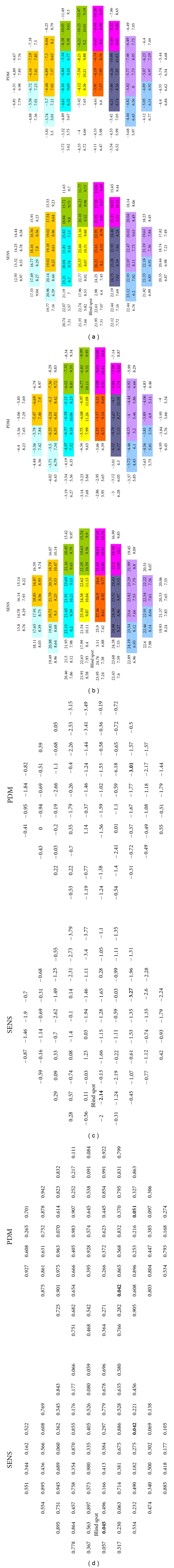
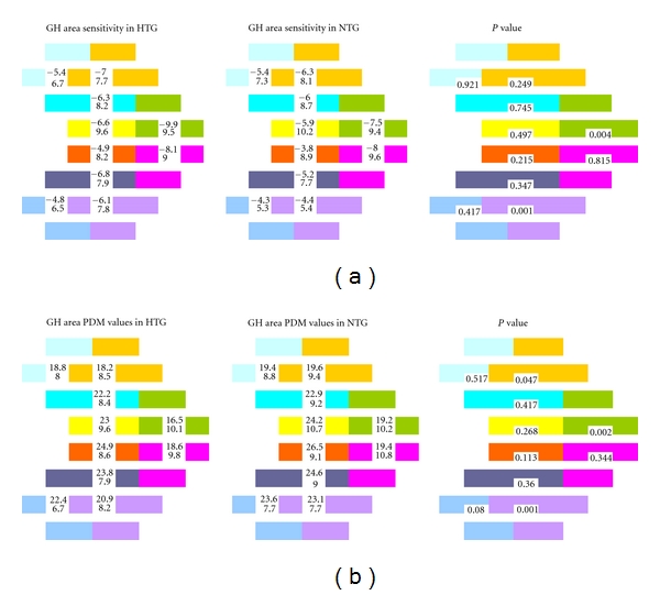
Purpose. To determine whether the patterns of visual field damage between high-tension glaucoma (HTG) and normal-tension glaucoma (NTG) are equivalent. Methods. In this retrospective cross-sectional study, fifty-one NTG and 57 HTG patients were recruited. For each recruited patient only the left eye was chosen. Glaucomatous patients had abnormal visual fields and/or glaucomatous changes at the optic nerve head. They were classified as HTG or NTG on the basis of intraocular pressure (IOP) measurements. Patients' visual fields were analyzed by using Humphrey Field Analyzer (HFA), program 30-2, full threshold. The visual field sensitivity values and the pattern deviation map values of the 72 tested points were considered. Then a pointwise analysis and an area analysis, based on the Glaucoma Hemifield test criteria, were performed, and a comparison between the two subgroups was made by Student's t test. Results. Between NTG and HTG, no significant difference was found pointwise for almost all the visual field points, except for two locations. One was under the blind spot, and the other was in the inferior hemifield around the twenty-degree position. When area analysis was considered, three areas showed a significantly different sensitivity between HTG and NTG. Conclusions. These data suggested that there was no relevant difference in the pointwise analysis between NTG and HTG; however, when visual field areas were compared, no difference in paracentral areas was found between NTG and HTG, but superior nasal step and inferior and superior scotomata showed to be deeper in HTG than in NTG.

摘要

目的。确定高眼压性青光眼(HTG)和正常眼压性青光眼(NTG)之间视野损害模式是否等同。方法。在这项回顾性横断面研究中,招募了51例NTG患者和57例HTG患者。对于每位招募的患者,仅选择左眼。青光眼患者视野异常和/或视神经乳头有青光眼性改变。根据眼压(IOP)测量结果将他们分为HTG或NTG。使用Humphrey视野分析仪(HFA)程序30 - 2全阈值分析患者的视野。考虑72个测试点的视野敏感度值和模式偏差图值。然后基于青光眼半视野测试标准进行逐点分析和区域分析,并通过学生t检验对两个亚组进行比较。结果。在NTG和HTG之间,几乎所有视野点逐点分析均未发现显著差异,但有两个位置除外。一个在盲点下方,另一个在20度位置周围的下半视野。当进行区域分析时,三个区域显示HTG和NTG之间敏感度有显著差异。结论。这些数据表明,NTG和HTG在逐点分析中没有相关差异;然而,当比较视野区域时,NTG和HTG在旁中心区域未发现差异,但HTG的鼻上阶梯以及下和上暗点比NTG更深。

https://cdn.ncbi.nlm.nih.gov/pmc/blobs/5ada/3306982/5353b839d00d/JOP2012-327326.002.jpg
https://cdn.ncbi.nlm.nih.gov/pmc/blobs/5ada/3306982/cc2910f00fbd/JOP2012-327326.001.jpg
https://cdn.ncbi.nlm.nih.gov/pmc/blobs/5ada/3306982/5353b839d00d/JOP2012-327326.002.jpg
https://cdn.ncbi.nlm.nih.gov/pmc/blobs/5ada/3306982/cc2910f00fbd/JOP2012-327326.001.jpg
https://cdn.ncbi.nlm.nih.gov/pmc/blobs/5ada/3306982/5353b839d00d/JOP2012-327326.002.jpg

相似文献

1
Visual field loss morphology in high- and normal-tension glaucoma.高眼压性青光眼和正常眼压性青光眼的视野缺损形态
J Ophthalmol. 2012;2012:327326. doi: 10.1155/2012/327326. Epub 2012 Feb 8.
2
Comparison of Superior and Inferior Visual Field Asymmetry Between Normal-tension and High-tension Glaucoma.正常眼压性青光眼与高眼压性青光眼上下视野不对称性的比较
J Glaucoma. 2021 Aug 1;30(8):648-655. doi: 10.1097/IJG.0000000000001872.
3
Morphological and functional differences between normal-tension and high-tension glaucoma.正常眼压性青光眼和高眼压性青光眼的形态和功能差异。
Acta Ophthalmol. 2013 Aug;91(5):e386-91. doi: 10.1111/aos.12061. Epub 2013 Feb 7.
4
Frequency doubling technology perimetry in open-angle glaucoma eyes with hemifield visual field damage: comparison of high-tension and normal-tension groups.倍频技术视野检查法在伴有半视野缺损的开角型青光眼眼中的应用:高眼压组与正常眼压组的比较
J Glaucoma. 2007 Jan;16(1):9-13. doi: 10.1097/01.ijg.0000243478.90403.84.
5
The Patterns of Visual Field Defects in Primary Angle-Closure Glaucoma Compared to High-Tension Glaucoma and Normal-Tension Glaucoma.原发性闭角型青光眼、高眼压型青光眼和正常眼压型青光眼视野缺损模式的比较。
Ophthalmic Res. 2023;66(1):940-948. doi: 10.1159/000530175. Epub 2023 Apr 14.
6
Comparison of normal- and high-tension glaucoma: nerve fiber layer and optic nerve head damage.正常眼压性青光眼与高眼压性青光眼的比较:神经纤维层及视神经乳头损害
Ophthalmologica. 2014;231(3):160-5. doi: 10.1159/000355326. Epub 2013 Dec 7.
7
Agreement between frequency doubling perimetry and static perimetry in eyes with high tension glaucoma and normal tension glaucoma.高眼压性青光眼和正常眼压性青光眼患者中倍频视野检查与静态视野检查之间的一致性
Br J Ophthalmol. 2003 May;87(5):604-8. doi: 10.1136/bjo.87.5.604.
8
Visual Field Defects in Young Patients With Open-angle Glaucoma: Comparison Between High-tension and Normal-tension Glaucoma.开角型青光眼年轻患者的视野缺损:高眼压性青光眼与正常眼压性青光眼的比较
J Glaucoma. 2017 Jun;26(6):541-547. doi: 10.1097/IJG.0000000000000667.
9
Normal tension vs high tension glaucoma: an - overview.正常眼压性青光眼与高眼压性青光眼:综述
Cesk Slov Oftalmol. 2019 Summer;75(2):55-60. doi: 10.31348/2019/2/1.
10
Visual field damage proximal to fixation in normal- and high-tension glaucoma eyes.正常眼压性青光眼和高眼压性青光眼患者注视点附近的视野损害
Jpn J Ophthalmol. 1995;39(3):274-83.

引用本文的文献

1
Visual field patterns in glaucoma: A systematic review.青光眼的视野模式:一项系统综述。
Saudi J Ophthalmol. 2024 Dec 26;38(4):306-315. doi: 10.4103/sjopt.sjopt_143_24. eCollection 2024 Oct-Dec.
2
Genome-wide meta-analysis identifies 22 loci for normal tension glaucoma with significant overlap with high tension glaucoma.全基因组荟萃分析确定了 22 个正常眼压性青光眼的位点,与高眼压性青光眼有显著重叠。
Nat Commun. 2024 Nov 17;15(1):9959. doi: 10.1038/s41467-024-54301-2.
3
Visualising structural and functional characteristics distinguishing between newly diagnosed high-tension and low-tension glaucoma patients.

本文引用的文献

1
Incorporating corneal pachymetry into the management of glaucoma.将角膜测厚法纳入青光眼的治疗中。
J Cataract Refract Surg. 2009 Sep;35(9):1623-8. doi: 10.1016/j.jcrs.2009.05.015.
2
Visual field and optic disc progression in patients with different types of optic disc damage: a longitudinal prospective study.不同类型视盘损伤患者的视野和视盘进展:一项纵向前瞻性研究。
Ophthalmology. 2003 Nov;110(11):2178-84. doi: 10.1016/S0161-6420(03)00801-7.
3
Optic nerve head morphologic characteristics in high-tension and normal-tension glaucoma.
可视化区分新发高压性和低压性青光眼患者的结构和功能特征。
Ophthalmic Physiol Opt. 2023 Jul;43(4):771-787. doi: 10.1111/opo.13129. Epub 2023 Mar 25.
4
Neuroinflammatory Mechanisms of Mitochondrial Dysfunction and Neurodegeneration in Glaucoma.青光眼中线粒体功能障碍和神经退行性变的神经炎症机制
J Ophthalmol. 2021 Apr 15;2021:4581909. doi: 10.1155/2021/4581909. eCollection 2021.
5
Spatial and Temporal Characteristics of Visual Field Progression in Glaucoma Assessed by Parallel Factor Analysis.通过平行因子分析评估青光眼视野进展的时空特征。
Korean J Ophthalmol. 2019 Jun;33(3):279-286. doi: 10.3341/kjo.2019.0004.
6
The nitric oxide-guanylate cyclase pathway and glaucoma.一氧化氮-鸟苷酸环化酶通路与青光眼。
Nitric Oxide. 2018 Jul 1;77:75-87. doi: 10.1016/j.niox.2018.04.010. Epub 2018 May 1.
7
Primary Open-Angle Glaucoma Genetics in African Americans.非裔美国人原发性开角型青光眼的遗传学
Curr Genet Med Rep. 2017 Dec;5(4):167-174. doi: 10.1007/s40142-017-0131-8. Epub 2017 Oct 11.
8
Plasma endothelin-1 and single nucleotide polymorphisms of as risk factors for normal tension glaucoma.血浆内皮素-1及[相关基因]单核苷酸多态性作为正常眼压性青光眼的危险因素
Mol Vis. 2016 Oct 20;22:1256-1266. eCollection 2016.
9
Vascular and autonomic dysregulation in primary open-angle glaucoma.原发性开角型青光眼中的血管和自主神经调节异常。
Curr Opin Ophthalmol. 2016 Mar;27(2):94-101. doi: 10.1097/ICU.0000000000000245.
10
Association of CAV1/CAV2 genomic variants with primary open-angle glaucoma overall and by gender and pattern of visual field loss.CAV1/CAV2 基因组变异与原发性开角型青光眼总体及性别和视野丧失模式的关联。
Ophthalmology. 2014 Feb;121(2):508-16. doi: 10.1016/j.ophtha.2013.09.012. Epub 2013 Oct 25.
高眼压性和正常眼压性青光眼的视神经乳头形态学特征
Arch Ophthalmol. 1999 Aug;117(8):1010-3. doi: 10.1001/archopht.117.8.1010.
4
Sector-based analysis of optic nerve head shape parameters and visual field indices in healthy and glaucomatous eyes.健康眼和青光眼眼中视神经乳头形状参数及视野指标的分区分析。
J Glaucoma. 1997 Dec;6(6):370-6.
5
Quantitative differences between the optic nerve head and peripapillary retina in low-tension and high-tension primary open-angle glaucoma.低眼压和高眼压原发性开角型青光眼视神经乳头与视乳头周围视网膜的定量差异
Am J Ophthalmol. 1997 Dec;124(6):805-13. doi: 10.1016/s0002-9394(14)71698-8.
6
Various glaucomatous optic nerve appearances: clinical correlations.各种青光眼性视神经表现:临床关联
Ophthalmology. 1996 Apr;103(4):640-9. doi: 10.1016/s0161-6420(96)30640-4.
7
Comparison of visual field defects between normal-tension and primary open-angle glaucoma in the late stage of the disease.正常眼压性青光眼与原发性开角型青光眼疾病晚期视野缺损的比较。
Graefes Arch Clin Exp Ophthalmol. 1995 Oct;233(10):610-6. doi: 10.1007/BF00185280.
8
Visual field defects in normal-tension and high-tension glaucoma.正常眼压性青光眼和高眼压性青光眼的视野缺损
Ophthalmology. 1993 Dec;100(12):1808-14. doi: 10.1016/s0161-6420(93)31394-1.
9
Focal and diffuse visual field defects: their relationship to intraocular pressure.局限性和弥漫性视野缺损:它们与眼压的关系。
Ophthalmic Surg. 1993 Aug;24(8):519-25.
10
Visual field defects in low-tension glaucoma. Comparison of defects in low-tension glaucoma and chronic open angle glaucoma.低眼压性青光眼的视野缺损。低眼压性青光眼与慢性开角型青光眼缺损的比较。
Arch Ophthalmol. 1982 Jul;100(7):1074-7. doi: 10.1001/archopht.1982.01030040052005.