Department of Neurology, University of Michigan, Ann Arbor, MI 48109-5622, USA.
Brain Res. 2012 May 25;1456:64-71. doi: 10.1016/j.brainres.2012.03.037. Epub 2012 Mar 23.
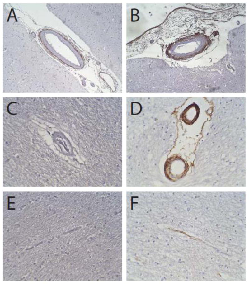
Arteries in cerebral autosomal dominant arteriopathy with subcortical infarcts and leukoencephalopathy (CADASIL) are susceptible to smooth muscle loss and fibrosis, but the molecular components underlying these dramatic vascular changes are not well characterized. The purpose of this study was to investigate the distribution of collagen isoforms in the cerebral vessels of North American CADASIL patients with classical NOTCH3 mutations. Expression of types I-VI collagen in brains obtained at autopsy from six CADASIL patients with cysteine-altering mutations in NOTCH3 was compared to control brain expression. We identified a consistent increase of types I, III, IV, and VI collagen in CADASIL brains. Strong accumulation of types I, III, IV and VI collagen was noted in all calibers of vessels, including small and medium-sized leptomeningeal arteries, small penetrating white matter arteries, and capillaries. Within leptomeningeal arteries, where we could define the three tunicae of each vessel, we found distinct collagen subtype distribution patterns in CADASIL. Types I and III collagen were largely found in either adventitial/medial or transmural locations. Type IV collagen was strictly intimal/medial. Type VI collagen was adventitial or adventitial/medial. Within the thickened penetrating arteries of CADASIL patients, all four collagens extended through most of the arterial wall. We observed increased staining of capillaries in CADASIL for types I, IV, and VI collagen. In conclusion, brain vascular collagen subtypes are increased in CADASIL in multiple layers of all sizes of arteries, with disease-specific changes most prominent in the tunica media and thickened small penetrating vessels. In diseased arteries, types I, III, and VI collagen spreads from an external location (adventitia) into the vascular media, while type IV collagen accumulates in an internal pattern (intima and media). These observations are consistent with a pathological role for collagen accumulation in the vascular media in CADASIL.
脑常染色体显性动脉病伴皮质下梗死和白质脑病(CADASIL)中的动脉容易发生平滑肌丧失和纤维化,但这些显著血管变化的分子成分尚未得到很好的描述。本研究的目的是研究具有经典 NOTCH3 突变的北美 CADASIL 患者脑血管中胶原异构体的分布。将 6 例 NOTCH3 中半胱氨酸改变突变的 CADASIL 患者尸检大脑中的胶原 I-VI 型表达与对照脑表达进行比较。我们发现 CADASIL 大脑中的胶原 I、III、IV 和 VI 型表达一致增加。在包括小和中等大小的脑膜动脉、小穿透性白质动脉和毛细血管在内的所有口径的血管中,都注意到胶原 I、III、IV 和 VI 型的强烈积累。在脑膜动脉中,我们可以定义每个血管的三个膜,我们发现 CADASIL 中存在不同的胶原亚型分布模式。胶原 I 和 III 型主要位于外膜/中膜或穿壁位置。IV 型胶原严格位于内膜/中膜。VI 型胶原位于外膜或外膜/中膜。在 CADASIL 患者增厚的穿透性动脉中,所有四种胶原都延伸到大部分动脉壁。我们观察到 CADASIL 患者的毛细血管中 I、IV 和 VI 型胶原的染色增加。总之,在所有大小的动脉的多层中,CADASIL 大脑血管胶原亚型增加,疾病特异性变化在中膜和增厚的小穿透性血管中最为明显。在患病的动脉中,胶原 I、III 和 VI 型从外部位置(外膜)扩散到血管中膜,而 IV 型胶原在内部模式(内膜和中膜)中积累。这些观察结果与 CADASIL 中血管中膜胶原积累的病理作用一致。