• 文献检索
  • 文档翻译
  • 深度研究
  • 学术资讯
  • Suppr Zotero 插件Zotero 插件
  • 邀请有礼
  • 套餐&价格
  • 历史记录
应用&插件
Suppr Zotero 插件Zotero 插件浏览器插件Mac 客户端Windows 客户端微信小程序
定价
高级版会员购买积分包购买API积分包
服务
文献检索文档翻译深度研究API 文档MCP 服务
关于我们
关于 Suppr公司介绍联系我们用户协议隐私条款
关注我们

Suppr 超能文献

核心技术专利:CN118964589B侵权必究
粤ICP备2023148730 号-1Suppr @ 2026

文献检索

告别复杂PubMed语法,用中文像聊天一样搜索,搜遍4000万医学文献。AI智能推荐,让科研检索更轻松。

立即免费搜索

文件翻译

保留排版,准确专业,支持PDF/Word/PPT等文件格式,支持 12+语言互译。

免费翻译文档

深度研究

AI帮你快速写综述,25分钟生成高质量综述,智能提取关键信息,辅助科研写作。

立即免费体验

经胸三维超声心动图的当前临床应用

Current clinical applications of transthoracic three-dimensional echocardiography.

作者信息

Badano Luigi P, Boccalini Francesca, Muraru Denisa, Bianco Lucia Dal, Peluso Diletta, Bellu Roberto, Zoppellaro Giacomo, Iliceto Sabino

机构信息

Department of Cardiac, Thoracic and Vascular Sciences, University of Padua, Padua, Italy.

出版信息

J Cardiovasc Ultrasound. 2012 Mar;20(1):1-22. doi: 10.4250/jcu.2012.20.1.1. Epub 2012 Mar 27.

DOI:10.4250/jcu.2012.20.1.1
PMID:22509433
原文链接:https://pmc.ncbi.nlm.nih.gov/articles/PMC3324722/
Abstract

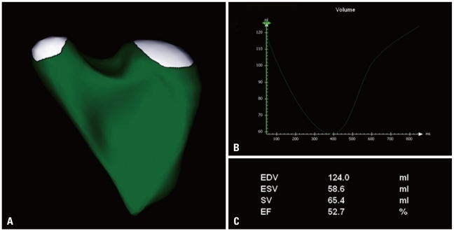
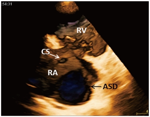
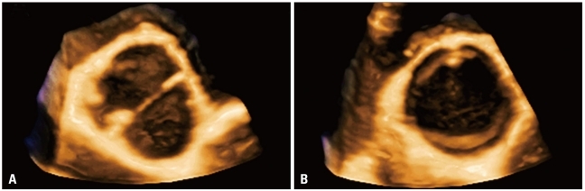
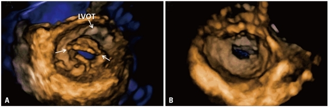
The advent of three-dimensional echocardiography (3DE) has significantly improved the impact of non-invasive imaging on our understanding and management of cardiac diseases in clinical practice. Transthoracic 3DE enables an easier, more accurate and reproducible interpretation of the complex cardiac anatomy, overcoming the intrinsic limitations of conventional echocardiography. The availability of unprecedented views of cardiac structures from any perspective in the beating heart provides valuable clinical information and new levels of confidence in diagnosing heart disease. One major advantage of the third dimension is the improvement in the accuracy and reproducibility of chamber volume measurement by eliminating geometric assumptions and errors caused by foreshortened views. Another benefit of 3DE is the realistic en face views of heart valves, enabling a better appreciation of the severity and mechanisms of valve diseases in a unique, noninvasive manner. The purpose of this review is to provide readers with an update on the current clinical applications of transthoracic 3DE, emphasizing the incremental benefits of 3DE over conventional two-dimensional echocardiography.

摘要

三维超声心动图(3DE)的出现显著提升了无创成像在临床实践中对我们理解和管理心脏疾病的影响。经胸三维超声心动图能够更轻松、准确且可重复地解读复杂的心脏解剖结构,克服了传统超声心动图的固有局限性。从跳动心脏的任何角度获取前所未有的心脏结构视图,为诊断心脏病提供了有价值的临床信息,并带来了新的信心水平。三维的一个主要优势是通过消除几何假设和因缩短视图导致的误差,提高了心腔容积测量的准确性和可重复性。三维超声心动图的另一个好处是能够获得心脏瓣膜逼真的正面视图,以独特的无创方式更好地评估瓣膜疾病的严重程度和机制。本综述的目的是向读者提供经胸三维超声心动图当前临床应用的最新情况,强调三维超声心动图相对于传统二维超声心动图的额外益处。

https://cdn.ncbi.nlm.nih.gov/pmc/blobs/64a1/3324722/af571fb500f3/jcu-20-1-g019.jpg
https://cdn.ncbi.nlm.nih.gov/pmc/blobs/64a1/3324722/a70544969c69/jcu-20-1-g001.jpg
https://cdn.ncbi.nlm.nih.gov/pmc/blobs/64a1/3324722/3fa6f4847ab1/jcu-20-1-g002.jpg
https://cdn.ncbi.nlm.nih.gov/pmc/blobs/64a1/3324722/8b02039f8887/jcu-20-1-g003.jpg
https://cdn.ncbi.nlm.nih.gov/pmc/blobs/64a1/3324722/ae405df9e412/jcu-20-1-g004.jpg
https://cdn.ncbi.nlm.nih.gov/pmc/blobs/64a1/3324722/7bddf4044ba0/jcu-20-1-g005.jpg
https://cdn.ncbi.nlm.nih.gov/pmc/blobs/64a1/3324722/f989196f15e1/jcu-20-1-g006.jpg
https://cdn.ncbi.nlm.nih.gov/pmc/blobs/64a1/3324722/2569ff538e91/jcu-20-1-g007.jpg
https://cdn.ncbi.nlm.nih.gov/pmc/blobs/64a1/3324722/94d8734f508b/jcu-20-1-g008.jpg
https://cdn.ncbi.nlm.nih.gov/pmc/blobs/64a1/3324722/1f24e539dc18/jcu-20-1-g009.jpg
https://cdn.ncbi.nlm.nih.gov/pmc/blobs/64a1/3324722/2e0e63bb2e25/jcu-20-1-g010.jpg
https://cdn.ncbi.nlm.nih.gov/pmc/blobs/64a1/3324722/cf218e047d8a/jcu-20-1-g011.jpg
https://cdn.ncbi.nlm.nih.gov/pmc/blobs/64a1/3324722/169fa2f2808c/jcu-20-1-g012.jpg
https://cdn.ncbi.nlm.nih.gov/pmc/blobs/64a1/3324722/c8294841aede/jcu-20-1-g013.jpg
https://cdn.ncbi.nlm.nih.gov/pmc/blobs/64a1/3324722/8c7379c6e0ac/jcu-20-1-g014.jpg
https://cdn.ncbi.nlm.nih.gov/pmc/blobs/64a1/3324722/70fa74731508/jcu-20-1-g015.jpg
https://cdn.ncbi.nlm.nih.gov/pmc/blobs/64a1/3324722/4941f0ef0953/jcu-20-1-g016.jpg
https://cdn.ncbi.nlm.nih.gov/pmc/blobs/64a1/3324722/fd83e8e0cbab/jcu-20-1-g017.jpg
https://cdn.ncbi.nlm.nih.gov/pmc/blobs/64a1/3324722/ccc46c585e3a/jcu-20-1-g018.jpg
https://cdn.ncbi.nlm.nih.gov/pmc/blobs/64a1/3324722/af571fb500f3/jcu-20-1-g019.jpg

相似文献

1
Current clinical applications of transthoracic three-dimensional echocardiography.经胸三维超声心动图的当前临床应用
J Cardiovasc Ultrasound. 2012 Mar;20(1):1-22. doi: 10.4250/jcu.2012.20.1.1. Epub 2012 Mar 27.
2
Current Clinical Applications of Three-Dimensional Echocardiography: When the Technique Makes the Difference.三维超声心动图的当前临床应用:当技术产生差异时。
Curr Cardiol Rep. 2016 Nov;18(11):109. doi: 10.1007/s11886-016-0787-9.
3
[Pathological diagnosis of conotruncal defects by transthoracic three-dimensional echocardiography].经胸三维超声心动图对圆锥干畸形的病理诊断
Zhonghua Er Ke Za Zhi. 2005 Sep;43(9):681-4.
4
European Association of Cardiovascular Imaging/Cardiovascular Imaging Department of the Brazilian Society of Cardiology recommendations for the use of cardiac imaging to assess and follow patients after heart transplantation.欧洲心血管影像协会/巴西心脏病学会心血管影像学部关于心脏移植后评估和随访患者使用心脏影像的建议。
Eur Heart J Cardiovasc Imaging. 2015 Sep;16(9):919-48. doi: 10.1093/ehjci/jev139. Epub 2015 Jul 2.
5
[Current clinical applications of three-dimensional echocardiography].[三维超声心动图的当前临床应用]
G Ital Cardiol (Rome). 2019 Dec;20(12):722-735. doi: 10.1714/3271.32381.
6
Valvular heart disease and 3-dimensional echocardiography: ready for prime-time?心脏瓣膜病与三维超声心动图:准备好进入黄金时代了吗?
Am J Cardiovasc Dis. 2012;2(4):279-84. Epub 2012 Oct 25.
7
Incremental value of the en face view of the tricuspid valve by two-dimensional and three-dimensional echocardiography for accurate identification of tricuspid valve leaflets.二维及三维超声心动图额面观在准确识别三尖瓣瓣叶中的增量价值。
J Am Soc Echocardiogr. 2014 Apr;27(4):376-84. doi: 10.1016/j.echo.2013.12.017. Epub 2014 Jan 31.
8
Mitral valve anatomy and function: new insights from three-dimensional echocardiography.二尖瓣解剖结构和功能:三维超声心动图的新见解。
J Cardiovasc Med (Hagerstown). 2013 Feb;14(2):91-9. doi: 10.2459/JCM.0b013e328356a577.
9
Diagnosis of mitral valve cleft using real-time 3-dimensional echocardiography.使用实时三维超声心动图诊断二尖瓣裂缺。
J Thorac Dis. 2017 Jan;9(1):159-165. doi: 10.21037/jtd.2017.01.21.
10
Tomographic three-dimensional echocardiographic determination of chamber size and systolic function in patients with left ventricular aneurysm: comparison to magnetic resonance imaging, cineventriculography, and two-dimensional echocardiography.断层三维超声心动图测定左心室室壁瘤患者的腔室大小和收缩功能:与磁共振成像、电影心室造影和二维超声心动图的比较
Circulation. 1997 Dec 16;96(12):4286-97. doi: 10.1161/01.cir.96.12.4286.

引用本文的文献

1
Comparison between Handheld Echocardiography and Cardiac Magnetic Resonance for Stroke Volume and Left Ventricular Ejection Fraction Quantification.手持式超声心动图与心脏磁共振成像在每搏输出量和左心室射血分数定量方面的比较
J Med Ultrasound. 2024 Aug 28;32(3):215-220. doi: 10.4103/jmu.jmu_52_23. eCollection 2024 Jul-Sep.
2
How Right is the Right Ventricle in Predicting Cardiac Mortality in Cardiac Failure: A 6-year Prospective Cohort Study.右心室在预测心力衰竭患者心脏死亡率方面的准确性如何:一项为期6年的前瞻性队列研究。
J Cardiovasc Echogr. 2024 Apr-Jun;34(2):50-56. doi: 10.4103/jcecho.jcecho_13_24. Epub 2024 Jun 28.
3

本文引用的文献

1
Feasibility and accuracy of 3DTEE versus CT for the evaluation of aortic valve annulus to left main ostium distance before transcatheter aortic valve implantation.经胸三维超声心动图与 CT 评估经导管主动脉瓣植入术前主动脉瓣环至左主干开口距离的可行性和准确性。
JACC Cardiovasc Imaging. 2012 Jun;5(6):579-88. doi: 10.1016/j.jcmg.2012.02.012.
2
Assessment of aortic valve complex by three-dimensional echocardiography: a framework for its effective application in clinical practice.三维超声心动图评估主动脉瓣复合体:有效应用于临床实践的框架。
Eur Heart J Cardiovasc Imaging. 2012 Jul;13(7):541-55. doi: 10.1093/ehjci/jes075. Epub 2012 Apr 19.
3
A Novel Automatic Algorithm to Support Lung Ultrasound Non-Expert Physicians in Interstitial Pneumonia Evaluation: A Single-Center Study.
一种支持非专业医生评估间质性肺炎的新型肺部超声自动算法:单中心研究
Diagnostics (Basel). 2024 Jan 10;14(2):155. doi: 10.3390/diagnostics14020155.
4
Position Statement on the Use of Myocardial Strain in Cardiology Routines by the Brazilian Society of Cardiology's Department Of Cardiovascular Imaging - 2023.《关于心肌应变在心血管影像学分会常规心脏病学中的应用的立场声明——2023》。
Arq Bras Cardiol. 2023 Dec;120(12):e20230646. doi: 10.36660/abc.20230646.
5
Evaluation of the image quality and validity of handheld echocardiography for stroke volume and left ventricular ejection fraction quantification: a method comparison study.手持式超声心动图评估脑卒中患者心排量和左心室射血分数的图像质量和有效性:方法比较研究。
Int J Cardiovasc Imaging. 2024 Jan;40(1):15-25. doi: 10.1007/s10554-023-02942-7. Epub 2023 Oct 10.
6
Soluble ST2 as a New Oxidative Stress and Inflammation Marker in Metabolic Syndrome.可溶性 ST2 作为代谢综合征中新的氧化应激和炎症标志物。
Int J Environ Res Public Health. 2023 Jan 31;20(3):2579. doi: 10.3390/ijerph20032579.
7
Multimodality Imaging of Sinus Venosus Atrial Septal Defect: A Challenging Diagnosis in Adults.静脉窦型房间隔缺损的多模态成像:成人中的一项具有挑战性的诊断
CASE (Phila). 2021 Dec 31;6(3):107-113. doi: 10.1016/j.case.2021.12.002. eCollection 2022 May.
8
Cardiooncology-dealing with modern drug treatment, long-term complications, and cancer survivorship.心脏肿瘤学——处理现代药物治疗、长期并发症和癌症生存问题。
Clin Exp Metastasis. 2021 Aug;38(4):361-371. doi: 10.1007/s10585-021-10106-x. Epub 2021 Jun 12.
9
The Role of Cardiac Magnetic Resonance in Evaluation of Idiopathic Ventricular Arrhythmia in Children.心脏磁共振成像在儿童特发性室性心律失常评估中的作用
J Clin Med. 2021 Mar 24;10(7):1335. doi: 10.3390/jcm10071335.
10
Usefulness of three-dimensional echocardiography for assessment of left and right ventricular volumes in children, verified by cardiac magnetic resonance. Can we overcome the discrepancy?经心脏磁共振验证的三维超声心动图在评估儿童左、右心室容积中的效用。我们能否克服这种差异?
Arch Med Sci. 2019 Apr 5;17(1):71-83. doi: 10.5114/aoms.2019.84215. eCollection 2021.
EAE/ASE recommendations for image acquisition and display using three-dimensional echocardiography.
欧洲超声心动图协会/美国超声心动图学会关于使用三维超声心动图进行图像采集和显示的建议。
Eur Heart J Cardiovasc Imaging. 2012 Jan;13(1):1-46. doi: 10.1093/ehjci/jer316.
4
Morphologic and functional remodeling of the right ventricle in pulmonary hypertension by real time three dimensional echocardiography.实时三维超声心动图对肺动脉高压右心室形态和功能的重构研究。
Am J Cardiol. 2012 Mar 15;109(6):906-13. doi: 10.1016/j.amjcard.2011.10.054. Epub 2011 Dec 22.
5
Sources of variation and bias in assessing left ventricular volumes and dyssynchrony using three-dimensional echocardiography.使用三维超声心动图评估左心室容积和不同步性的变异性和偏倚来源。
Int J Cardiovasc Imaging. 2012 Aug;28(6):1357-68. doi: 10.1007/s10554-011-9985-0. Epub 2011 Nov 27.
6
Valvular heart disease. The value of 3-dimensional echocardiography.瓣膜性心脏病。三维超声心动图的价值。
J Am Coll Cardiol. 2011 Nov 1;58(19):1933-44. doi: 10.1016/j.jacc.2011.07.035.
7
EAE/ASE recommendations for the use of echocardiography in new transcatheter interventions for valvular heart disease.欧洲心胸外科学会(EAE)/美国超声心动图学会(ASE)关于超声心动图在心脏瓣膜病新经导管介入治疗中应用的建议。
Eur J Echocardiogr. 2011 Aug;12(8):557-84. doi: 10.1093/ejechocard/jer086.
8
Evaluation of the left atrial appendage with real-time 3-dimensional transesophageal echocardiography: implications for catheter-based left atrial appendage closure.实时三维经食管超声心动图评价左心耳:经导管左心耳封堵的意义。
Circ Cardiovasc Imaging. 2011 Sep;4(5):514-23. doi: 10.1161/CIRCIMAGING.111.963892. Epub 2011 Jul 7.
9
A three-dimensional insight into the complexity of flow convergence in mitral regurgitation: adjunctive benefit of anatomic regurgitant orifice area.深入了解二尖瓣反流中血流汇聚的复杂性:解剖性反流口面积的附加获益。
Am J Physiol Heart Circ Physiol. 2011 Sep;301(3):H1015-24. doi: 10.1152/ajpheart.00275.2011. Epub 2011 Jun 10.
10
Recommendations of the European Association of Echocardiography: how to use echo-Doppler in clinical trials: different modalities for different purposes.欧洲超声心动图协会的建议:如何在临床试验中使用超声多普勒:不同目的采用不同模式。
Eur J Echocardiogr. 2011 May;12(5):339-53. doi: 10.1093/ejechocard/jer051.