Program in Cancer Biology, Stanford University, Stanford, CA 94305, USA.
Curr Biol. 2012 May 8;22(9):837-42. doi: 10.1016/j.cub.2012.03.037. Epub 2012 Apr 19.
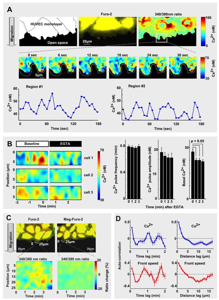
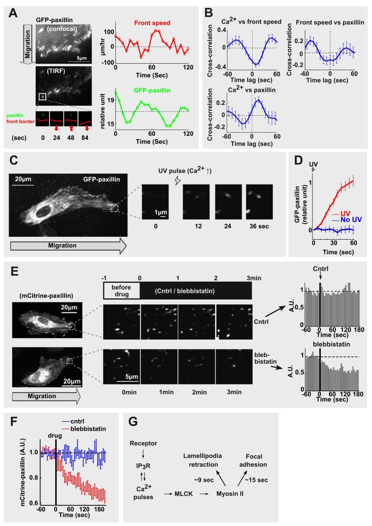
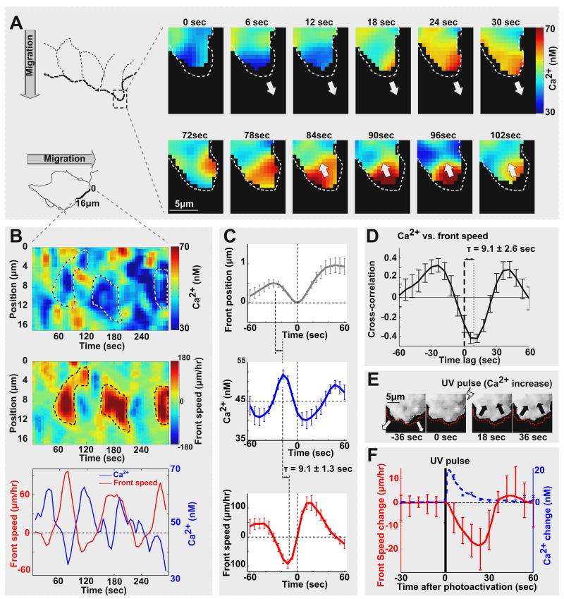
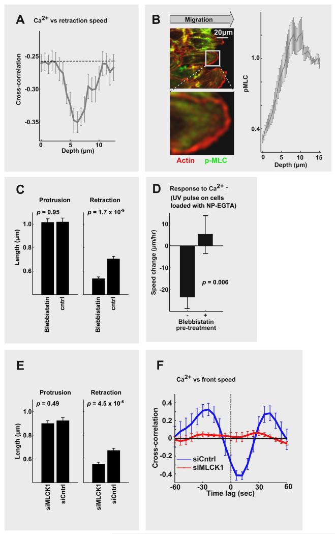
Ca(2+) signals regulate polarization, speed, and turning of migrating cells. However, the molecular mechanism by which Ca(2+) acts on moving cells is not understood. Here we show that local Ca(2+) pulses along the front of migrating human endothelial cells trigger cycles of retraction of local lamellipodia and, concomitantly, strengthen local adhesion to the extracellular matrix. These Ca(2+) release pulses had small amplitudes and diameters and were triggered repetitively near the leading plasma membrane with only little coordination between different regions. We show that each Ca(2+) pulse triggers contraction of actin filaments by activating myosin light-chain kinase and myosin II behind the leading edge. The cyclic force generated by myosin II operates locally, causing a partial retraction of the nearby protruding lamellipodia membrane and a strengthening of paxillin-based focal adhesion within the same lamellipodia. Photo release of Ca(2+) demonstrated a direct role of Ca(2+) in triggering local retraction and adhesion. Together, our study suggests that spatial sensing, forward movement, turning, and chemotaxis are in part controlled by confined Ca(2+) pulses that promote local lamellipodia retraction and adhesion cycles along the leading edge of moving cells.
钙离子信号调节迁移细胞的极化、速度和转向。然而,钙离子作用于运动细胞的分子机制尚不清楚。在这里,我们显示了局部钙离子脉冲沿着迁移的人内皮细胞的前缘引发局部片状伪足的收缩循环,同时,加强了与细胞外基质的局部黏附。这些钙离子释放脉冲幅度和直径都很小,并且在靠近前导质膜的位置反复触发,不同区域之间只有很少的协调。我们表明,每个钙离子脉冲通过激活肌球蛋白轻链激酶和肌球蛋白 II 来触发前缘后面的肌动蛋白丝的收缩。肌球蛋白 II 产生的循环力在局部起作用,导致附近突出的片状伪足膜的部分缩回,并在同一片状伪足内加强了基于桩蛋白的黏附。钙离子的光释放证明了钙离子在触发局部缩回和黏附中的直接作用。总之,我们的研究表明,空间感知、向前运动、转向和趋化性部分受到限制的钙离子脉冲的控制,这些脉冲促进了运动细胞前缘的局部片状伪足缩回和黏附循环。